Gulfie Dentists Students
Evergreen Performance Test
Exams in alternate weeks to test your level of preparation.

Test of the Week
Scheduled for alternate Week
Test of the Week
PERFORMANCE TEST
Quiz-summary
0 of 150 questions completed
Questions:
- 1
- 2
- 3
- 4
- 5
- 6
- 7
- 8
- 9
- 10
- 11
- 12
- 13
- 14
- 15
- 16
- 17
- 18
- 19
- 20
- 21
- 22
- 23
- 24
- 25
- 26
- 27
- 28
- 29
- 30
- 31
- 32
- 33
- 34
- 35
- 36
- 37
- 38
- 39
- 40
- 41
- 42
- 43
- 44
- 45
- 46
- 47
- 48
- 49
- 50
- 51
- 52
- 53
- 54
- 55
- 56
- 57
- 58
- 59
- 60
- 61
- 62
- 63
- 64
- 65
- 66
- 67
- 68
- 69
- 70
- 71
- 72
- 73
- 74
- 75
- 76
- 77
- 78
- 79
- 80
- 81
- 82
- 83
- 84
- 85
- 86
- 87
- 88
- 89
- 90
- 91
- 92
- 93
- 94
- 95
- 96
- 97
- 98
- 99
- 100
- 101
- 102
- 103
- 104
- 105
- 106
- 107
- 108
- 109
- 110
- 111
- 112
- 113
- 114
- 115
- 116
- 117
- 118
- 119
- 120
- 121
- 122
- 123
- 124
- 125
- 126
- 127
- 128
- 129
- 130
- 131
- 132
- 133
- 134
- 135
- 136
- 137
- 138
- 139
- 140
- 141
- 142
- 143
- 144
- 145
- 146
- 147
- 148
- 149
- 150
Information
Click start this Evergreen Performance Test.
You have already completed the quiz before. Hence you can not start it again.
Quiz is loading...
You must sign in or sign up to start the quiz.
You have to finish following quiz, to start this quiz:
Results
0 of 150 questions answered correctly
Time has elapsed
You have reached 0 of 0 points, (0)
| Average score |
|
| Your score |
|
Categories
- KDLE 0%
| Pos. | Name | Entered on | Points | Result |
|---|---|---|---|---|
| Table is loading | ||||
| No data available | ||||
- 1
- 2
- 3
- 4
- 5
- 6
- 7
- 8
- 9
- 10
- 11
- 12
- 13
- 14
- 15
- 16
- 17
- 18
- 19
- 20
- 21
- 22
- 23
- 24
- 25
- 26
- 27
- 28
- 29
- 30
- 31
- 32
- 33
- 34
- 35
- 36
- 37
- 38
- 39
- 40
- 41
- 42
- 43
- 44
- 45
- 46
- 47
- 48
- 49
- 50
- 51
- 52
- 53
- 54
- 55
- 56
- 57
- 58
- 59
- 60
- 61
- 62
- 63
- 64
- 65
- 66
- 67
- 68
- 69
- 70
- 71
- 72
- 73
- 74
- 75
- 76
- 77
- 78
- 79
- 80
- 81
- 82
- 83
- 84
- 85
- 86
- 87
- 88
- 89
- 90
- 91
- 92
- 93
- 94
- 95
- 96
- 97
- 98
- 99
- 100
- 101
- 102
- 103
- 104
- 105
- 106
- 107
- 108
- 109
- 110
- 111
- 112
- 113
- 114
- 115
- 116
- 117
- 118
- 119
- 120
- 121
- 122
- 123
- 124
- 125
- 126
- 127
- 128
- 129
- 130
- 131
- 132
- 133
- 134
- 135
- 136
- 137
- 138
- 139
- 140
- 141
- 142
- 143
- 144
- 145
- 146
- 147
- 148
- 149
- 150
- Answered
- Review
-
Question 1 of 150
1. Question
A newborn presents with a defect characterized by a lack of continuity of the roof of the mouth involving part or the entire length of the palate, forming a fissure extending in the anteroposterior direction. Which condition does this description represent?
Correct
ANSWER
Congenital cleft palateOTHER OPTIONS
● Not applicableSYNOPSIS
● Congenital cleft palate results from failure of fusion of the palatal shelves during embryonic development.
● The defect may involve the soft palate alone or extend through the hard palate and alveolus, producing a fissure that runs anteroposteriorly.
● Patients with cleft palate commonly experience feeding difficulties, speech abnormalities, and recurrent middle ear infections, necessitating multidisciplinary management.REFERENCE
Peterson’s Principles of Oral and Maxillofacial Surgery – 7th Edition.Incorrect
ANSWER
Congenital cleft palateOTHER OPTIONS
● Not applicableSYNOPSIS
● Congenital cleft palate results from failure of fusion of the palatal shelves during embryonic development.
● The defect may involve the soft palate alone or extend through the hard palate and alveolus, producing a fissure that runs anteroposteriorly.
● Patients with cleft palate commonly experience feeding difficulties, speech abnormalities, and recurrent middle ear infections, necessitating multidisciplinary management.REFERENCE
Peterson’s Principles of Oral and Maxillofacial Surgery – 7th Edition. -
Question 2 of 150
2. Question
A 30-year-old woman presents with fatigue, pallor, angular cheilitis, and glossitis. Laboratory investigations reveal a decreased hemoglobin level with reduced mean corpuscular volume (MCV) and mean corpuscular hemoglobin (MCH). Which type of anemia is most consistent with these findings?
Correct
ANSWER
Microcytic anemiaOTHER OPTIONS
● Macrocytic anemia – Characterized by enlarged red blood cells and is commonly associated with vitamin B₁₂ or folate deficiency.
● Normocytic anemia – Characterized by normal-sized red blood cells and is commonly seen in acute blood loss, anemia of chronic disease, or aplastic anemia.SYNOPSIS
● Iron deficiency anemia is the most common nutritional anemia and is characterized by microcytic, hypochromic red blood cells.
● Oral manifestations include atrophic glossitis, angular cheilitis, mucosal pallor, and burning mouth symptoms.
● Diagnosis is based on complete blood count, reduced MCV and MCH, and iron studies demonstrating low serum ferritin and serum iron.REFERENCE
Little and Falace’s Dental Management of the Medically Compromised Patient – 10th EditionIncorrect
ANSWER
Microcytic anemiaOTHER OPTIONS
● Macrocytic anemia – Characterized by enlarged red blood cells and is commonly associated with vitamin B₁₂ or folate deficiency.
● Normocytic anemia – Characterized by normal-sized red blood cells and is commonly seen in acute blood loss, anemia of chronic disease, or aplastic anemia.SYNOPSIS
● Iron deficiency anemia is the most common nutritional anemia and is characterized by microcytic, hypochromic red blood cells.
● Oral manifestations include atrophic glossitis, angular cheilitis, mucosal pallor, and burning mouth symptoms.
● Diagnosis is based on complete blood count, reduced MCV and MCH, and iron studies demonstrating low serum ferritin and serum iron.REFERENCE
Little and Falace’s Dental Management of the Medically Compromised Patient – 10th Edition -
Question 3 of 150
3. Question
A 62-year-old patient with chronic liver cirrhosis and congestive heart failure is scheduled for a dental extraction under local anesthesia. Which of the following systemic conditions can reduce the hepatic metabolism and clearance of lignocaine, directly or indirectly thereby increasing the risk of local anesthetic toxicity?
Correct
ANSWER
All of the aboveOTHER OPTIONS
● NilSYNOPSIS
● Lignocaine is primarily metabolized in the liver; therefore, conditions affecting hepatic function or hepatic blood flow reduce its clearance.
● Liver cirrhosis directly impairs hepatic metabolism, while hypotension and congestive heart failure decrease hepatic perfusion, resulting in slower drug metabolism.
● Patients with these conditions are at an increased risk of local anesthetic toxicity, and dose reduction with careful monitoring is recommended.REFERENCE
Malamed SF. Handbook of Local Anesthesia – 7th EditionIncorrect
ANSWER
All of the aboveOTHER OPTIONS
● NilSYNOPSIS
● Lignocaine is primarily metabolized in the liver; therefore, conditions affecting hepatic function or hepatic blood flow reduce its clearance.
● Liver cirrhosis directly impairs hepatic metabolism, while hypotension and congestive heart failure decrease hepatic perfusion, resulting in slower drug metabolism.
● Patients with these conditions are at an increased risk of local anesthetic toxicity, and dose reduction with careful monitoring is recommended.REFERENCE
Malamed SF. Handbook of Local Anesthesia – 7th Edition -
Question 4 of 150
4. Question
Which systemic condition is a contraindication to the use of epinephrine-impregnated gingival retraction cords because of the risk of adverse cardiovascular effects?
Correct
ANSWER
HypertensionOTHER OPTIONS
● Asthma – Epinephrine-impregnated retraction cords are not specifically contraindicated in asthma, although caution may be required in patients with sulfite sensitivity.
● Crohn disease – There is no direct contraindication to the use of epinephrine-containing gingival retraction cords in patients with Crohn disease.
● Infective endocarditis – The use of epinephrine retraction cords does not increase the risk of infective endocarditis; management focuses on appropriate antibiotic prophylaxis when indicated.SYNOPSIS
● Epinephrine-impregnated gingival retraction cords can result in systemic absorption of epinephrine, leading to increased heart rate and blood pressure.
● Their use should be avoided or used with extreme caution in patients with uncontrolled hypertension and significant cardiovascular disease.
● Alternative retraction agents, such as aluminum chloride or ferric sulfate, are preferred in medically compromised patients at risk of adverse cardiovascular effects.REFERENCE
Rosenstiel SF, Land MF, Fujimoto J. Contemporary Fixed Prosthodontics – 6th EditionIncorrect
ANSWER
HypertensionOTHER OPTIONS
● Asthma – Epinephrine-impregnated retraction cords are not specifically contraindicated in asthma, although caution may be required in patients with sulfite sensitivity.
● Crohn disease – There is no direct contraindication to the use of epinephrine-containing gingival retraction cords in patients with Crohn disease.
● Infective endocarditis – The use of epinephrine retraction cords does not increase the risk of infective endocarditis; management focuses on appropriate antibiotic prophylaxis when indicated.SYNOPSIS
● Epinephrine-impregnated gingival retraction cords can result in systemic absorption of epinephrine, leading to increased heart rate and blood pressure.
● Their use should be avoided or used with extreme caution in patients with uncontrolled hypertension and significant cardiovascular disease.
● Alternative retraction agents, such as aluminum chloride or ferric sulfate, are preferred in medically compromised patients at risk of adverse cardiovascular effects.REFERENCE
Rosenstiel SF, Land MF, Fujimoto J. Contemporary Fixed Prosthodontics – 6th Edition -
Question 5 of 150
5. Question
A 55-year-old patient with a history of liver cirrhosis is scheduled for a dental extraction. Which laboratory investigation is the most important to evaluate the patient’s bleeding risk before performing the procedure?
Correct
ANSWER
Prothrombin timeOTHER OPTIONS
● D-dimer – Used primarily to evaluate thromboembolic disorders such as deep vein thrombosis or disseminated intravascular coagulation and is not routinely indicated before dental extraction in patients with liver disease.
● Serum calcium level – Assesses calcium metabolism but does not evaluate the patient’s bleeding tendency.
● Blood urea nitrogen (BUN) – Reflects renal function and is not used to assess coagulation status in patients with liver disease.SYNOPSIS
● The liver synthesizes most coagulation factors; therefore, liver disease may result in impaired coagulation and an increased risk of bleeding.
● Prothrombin time (PT) and International Normalized Ratio (INR) are the primary investigations used to assess coagulation status before invasive dental procedures in patients with liver disease.
● Additional investigations such as a complete blood count, including platelet count, and liver function tests may also be indicated depending on the severity of hepatic impairment.REFERENCE
Little and Falace’s Dental Management of the Medically Compromised Patient – 10th EditionIncorrect
ANSWER
Prothrombin timeOTHER OPTIONS
● D-dimer – Used primarily to evaluate thromboembolic disorders such as deep vein thrombosis or disseminated intravascular coagulation and is not routinely indicated before dental extraction in patients with liver disease.
● Serum calcium level – Assesses calcium metabolism but does not evaluate the patient’s bleeding tendency.
● Blood urea nitrogen (BUN) – Reflects renal function and is not used to assess coagulation status in patients with liver disease.SYNOPSIS
● The liver synthesizes most coagulation factors; therefore, liver disease may result in impaired coagulation and an increased risk of bleeding.
● Prothrombin time (PT) and International Normalized Ratio (INR) are the primary investigations used to assess coagulation status before invasive dental procedures in patients with liver disease.
● Additional investigations such as a complete blood count, including platelet count, and liver function tests may also be indicated depending on the severity of hepatic impairment.REFERENCE
Little and Falace’s Dental Management of the Medically Compromised Patient – 10th Edition -
Question 6 of 150
6. Question
A patient develops an acute bronchial asthma attack during dental treatment. Which of the following provides the most rapid relief of bronchospasm?
Correct
ANSWER
Short-acting bronchodilatorOTHER OPTIONS
● Oxygen and IV corticosteroids – Oxygen is supportive, and corticosteroids have a delayed onset of action; they do not provide immediate bronchodilation.
● Oxygen – Corrects hypoxemia but does not relieve bronchospasm.
● Epinephrine injection – Reserved for severe, life-threatening asthma unresponsive to inhaled bronchodilators or when anaphylaxis is suspected; it is not the first-line treatment for a typical acute asthma attack.SYNOPSIS
● In an acute asthma attack, an inhaled short-acting β₂-agonist (SABA) such as salbutamol is the first-line treatment because of its rapid bronchodilatory effect.
● Supplemental oxygen should be administered if hypoxemia is present, while systemic corticosteroids are used to reduce airway inflammation but have a delayed onset of action.
● Intramuscular epinephrine is reserved for severe refractory bronchospasm or asthma associated with anaphylaxis.REFERENCE
Malamed SF. Medical Emergencies in the Dental Office – 8th EditionIncorrect
ANSWER
Short-acting bronchodilatorOTHER OPTIONS
● Oxygen and IV corticosteroids – Oxygen is supportive, and corticosteroids have a delayed onset of action; they do not provide immediate bronchodilation.
● Oxygen – Corrects hypoxemia but does not relieve bronchospasm.
● Epinephrine injection – Reserved for severe, life-threatening asthma unresponsive to inhaled bronchodilators or when anaphylaxis is suspected; it is not the first-line treatment for a typical acute asthma attack.SYNOPSIS
● In an acute asthma attack, an inhaled short-acting β₂-agonist (SABA) such as salbutamol is the first-line treatment because of its rapid bronchodilatory effect.
● Supplemental oxygen should be administered if hypoxemia is present, while systemic corticosteroids are used to reduce airway inflammation but have a delayed onset of action.
● Intramuscular epinephrine is reserved for severe refractory bronchospasm or asthma associated with anaphylaxis.REFERENCE
Malamed SF. Medical Emergencies in the Dental Office – 8th Edition -
Question 7 of 150
7. Question
What is the average incubation period of hepatitis B virus infection?
Correct
ANSWER
90 daysOTHER OPTIONS
● Not applicableSYNOPSIS
● The average incubation period for hepatitis B virus (HBV) infection is 90 days (about 3 months), with a total range of 30 to 180 days from exposure to the onset of symptoms.
● During the incubation period, infected individuals may be asymptomatic but can still transmit the virus.
● Knowledge of the incubation period is important when evaluating occupational exposures and planning post-exposure prophylaxis.REFERENCE
Little and Falace’s Dental Management of the Medically Compromised Patient – 10th EditionIncorrect
ANSWER
90 daysOTHER OPTIONS
● Not applicableSYNOPSIS
● The average incubation period for hepatitis B virus (HBV) infection is 90 days (about 3 months), with a total range of 30 to 180 days from exposure to the onset of symptoms.
● During the incubation period, infected individuals may be asymptomatic but can still transmit the virus.
● Knowledge of the incubation period is important when evaluating occupational exposures and planning post-exposure prophylaxis.REFERENCE
Little and Falace’s Dental Management of the Medically Compromised Patient – 10th Edition -
Question 8 of 150
8. Question
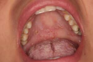
A patient with a history of inflammatory bowel disease presents with multiple painless snail-track ulcerations on the soft palate, consistent with pyostomatitis vegetans. What is the most appropriate management?
Correct
ANSWER
Systemic drugsOTHER OPTIONS
● IV diazepam – Diazepam has no role in the management of oral lesions associated with inflammatory bowel disease.
● Adrenaline – Adrenaline is indicated for anaphylaxis and certain medical emergencies, not for inflammatory oral lesions.SYNOPSIS
● Snail-track lesions of the oral mucosa are characteristic of pyostomatitis vegetans, an oral manifestation strongly associated with inflammatory bowel disease, particularly ulcerative colitis.
● Management primarily involves treating the underlying inflammatory bowel disease with systemic medications such as corticosteroids or immunosuppressive agents.
● Improvement of the systemic disease usually results in resolution of the oral lesions, while topical corticosteroids may be used as adjunctive therapy for symptomatic relief.REFERENCE
Burket’s Oral Medicine – 13th EditionIncorrect
ANSWER
Systemic drugsOTHER OPTIONS
● IV diazepam – Diazepam has no role in the management of oral lesions associated with inflammatory bowel disease.
● Adrenaline – Adrenaline is indicated for anaphylaxis and certain medical emergencies, not for inflammatory oral lesions.SYNOPSIS
● Snail-track lesions of the oral mucosa are characteristic of pyostomatitis vegetans, an oral manifestation strongly associated with inflammatory bowel disease, particularly ulcerative colitis.
● Management primarily involves treating the underlying inflammatory bowel disease with systemic medications such as corticosteroids or immunosuppressive agents.
● Improvement of the systemic disease usually results in resolution of the oral lesions, while topical corticosteroids may be used as adjunctive therapy for symptomatic relief.REFERENCE
Burket’s Oral Medicine – 13th Edition -
Question 9 of 150
9. Question
A 42-year-old patient with acute leukemia requires a dental extraction. Recent laboratory investigations reveal an absolute neutrophil count (ANC) of 1,700 cells/mm³, and the platelet count is within the normal range. What is the most appropriate dental management
Correct
ANSWER
Proceed with treatment using prophylactic antibioticsOTHER OPTIONS
● Postpone the dental procedure – An ANC of 1,700 cells/mm³ is above the threshold at which routine dental treatment must be deferred, provided the patient is otherwise medically stable.
● Administer a platelet transfusion before treatment – Platelet transfusion is based on the platelet count, not the neutrophil count, and is unnecessary when platelet levels are adequate.
● Proceed with treatment without special precautions – Patients with leukemia and an ANC between 1,000 and 2,000 cells/mm³ are at increased risk of infection; prophylactic antibiotics should be considered in consultation with the treating physician.SYNOPSIS
● The absolute neutrophil count (ANC) is an important indicator of infection risk before invasive dental procedures in patients with leukemia.
● Patients with an ANC >2,000 cells/mm³ generally do not require antibiotic prophylaxis, whereas those with an ANC between 1,000 and 2,000 cells/mm³ may require prophylactic antibiotics depending on the procedure and physician recommendations.
● Elective invasive dental procedures are generally deferred when the ANC is below 1,000 cells/mm³ because of the significantly increased risk of infection.REFERENCE
Little and Falace’s Dental Management of the Medically Compromised Patient – 10th EditionIncorrect
ANSWER
Proceed with treatment using prophylactic antibioticsOTHER OPTIONS
● Postpone the dental procedure – An ANC of 1,700 cells/mm³ is above the threshold at which routine dental treatment must be deferred, provided the patient is otherwise medically stable.
● Administer a platelet transfusion before treatment – Platelet transfusion is based on the platelet count, not the neutrophil count, and is unnecessary when platelet levels are adequate.
● Proceed with treatment without special precautions – Patients with leukemia and an ANC between 1,000 and 2,000 cells/mm³ are at increased risk of infection; prophylactic antibiotics should be considered in consultation with the treating physician.SYNOPSIS
● The absolute neutrophil count (ANC) is an important indicator of infection risk before invasive dental procedures in patients with leukemia.
● Patients with an ANC >2,000 cells/mm³ generally do not require antibiotic prophylaxis, whereas those with an ANC between 1,000 and 2,000 cells/mm³ may require prophylactic antibiotics depending on the procedure and physician recommendations.
● Elective invasive dental procedures are generally deferred when the ANC is below 1,000 cells/mm³ because of the significantly increased risk of infection.REFERENCE
Little and Falace’s Dental Management of the Medically Compromised Patient – 10th Edition -
Question 10 of 150
10. Question
Which systemic condition is considered a contraindication to administering an inferior alveolar nerve block without prior factor replacement therapy because of the risk of deep tissue hematoma?
Correct
ANSWER
Hemophilia AOTHER OPTIONS
● Thrombocytopenia – Inferior alveolar nerve block is contraindicated only in patients with severe thrombocytopenia or uncontrolled bleeding risk; the decision depends on the platelet count rather than the diagnosis alone.
● Hypoprothrombinemia – Patients require correction of the coagulation defect before invasive procedures. It is not an absolute contraindication in all cases.
● Von Willebrand disease – Inferior alveolar nerve block may be performed after appropriate medical management (e.g., desmopressin or factor replacement), depending on the severity of the disease.SYNOPSIS
● Inferior alveolar nerve block carries a risk of deep tissue hemorrhage that may lead to airway compromise in patients with severe coagulation disorders.
● In untreated severe hemophilia, an inferior alveolar nerve block should not be administered unless adequate clotting factor replacement has been provided in consultation with the patient’s hematologist.
● Alternative techniques, such as infiltration, intraligamentary, or intraosseous anesthesia, may be preferred when appropriate.REFERENCE
Little and Falace’s Dental Management of the Medically Compromised Patient – 10th EditionIncorrect
ANSWER
Hemophilia AOTHER OPTIONS
● Thrombocytopenia – Inferior alveolar nerve block is contraindicated only in patients with severe thrombocytopenia or uncontrolled bleeding risk; the decision depends on the platelet count rather than the diagnosis alone.
● Hypoprothrombinemia – Patients require correction of the coagulation defect before invasive procedures. It is not an absolute contraindication in all cases.
● Von Willebrand disease – Inferior alveolar nerve block may be performed after appropriate medical management (e.g., desmopressin or factor replacement), depending on the severity of the disease.SYNOPSIS
● Inferior alveolar nerve block carries a risk of deep tissue hemorrhage that may lead to airway compromise in patients with severe coagulation disorders.
● In untreated severe hemophilia, an inferior alveolar nerve block should not be administered unless adequate clotting factor replacement has been provided in consultation with the patient’s hematologist.
● Alternative techniques, such as infiltration, intraligamentary, or intraosseous anesthesia, may be preferred when appropriate.REFERENCE
Little and Falace’s Dental Management of the Medically Compromised Patient – 10th Edition -
Question 11 of 150
11. Question
A 58-year-old overweight man with a history of hypertension and dyslipidemia develops severe central chest pain during a dental procedure. The pain radiates to the right shoulder and arm, persists for more than 15 minutes, and does not subside after administration of sublingual nitroglycerin. Which of the following is the most likely diagnosis?
Correct
ANSWER
Myocardial infarctionOTHER OPTIONS
● Stable angina pectoris – Stable angina is usually precipitated by exertion or stress, lasts less than 10–15 minutes, and is relieved by rest or sublingual nitroglycerin.
● Unstable angina pectoris – Unstable angina may occur at rest and is more severe than stable angina, but the pain often responds at least partially to nitroglycerin and does not cause myocardial necrosis. Persistent pain unrelieved by nitroglycerin is more suggestive of myocardial infarction.
● Congestive heart failure – Congestive heart failure typically presents with dyspnea, orthopnea, fatigue, and peripheral edema rather than prolonged crushing chest pain.SYNOPSIS
● Myocardial infarction (MI) is characterized by prolonged, severe chest pain lasting more than 15–20 minutes, often radiating to the shoulder, arm, neck, or jaw, and not relieved by rest or nitroglycerin.
● Major risk factors include hypertension, dyslipidemia, obesity, diabetes mellitus, and smoking.
● During a dental procedure, suspected MI requires immediate termination of treatment, activation of emergency medical services, administration of aspirin (if not contraindicated), oxygen when indicated, and continuous monitoring until advanced medical care arrives.REFERENCE
Malamed SF. Medical Emergencies in the Dental Office – 8th EditionIncorrect
ANSWER
Myocardial infarctionOTHER OPTIONS
● Stable angina pectoris – Stable angina is usually precipitated by exertion or stress, lasts less than 10–15 minutes, and is relieved by rest or sublingual nitroglycerin.
● Unstable angina pectoris – Unstable angina may occur at rest and is more severe than stable angina, but the pain often responds at least partially to nitroglycerin and does not cause myocardial necrosis. Persistent pain unrelieved by nitroglycerin is more suggestive of myocardial infarction.
● Congestive heart failure – Congestive heart failure typically presents with dyspnea, orthopnea, fatigue, and peripheral edema rather than prolonged crushing chest pain.SYNOPSIS
● Myocardial infarction (MI) is characterized by prolonged, severe chest pain lasting more than 15–20 minutes, often radiating to the shoulder, arm, neck, or jaw, and not relieved by rest or nitroglycerin.
● Major risk factors include hypertension, dyslipidemia, obesity, diabetes mellitus, and smoking.
● During a dental procedure, suspected MI requires immediate termination of treatment, activation of emergency medical services, administration of aspirin (if not contraindicated), oxygen when indicated, and continuous monitoring until advanced medical care arrives.REFERENCE
Malamed SF. Medical Emergencies in the Dental Office – 8th Edition -
Question 12 of 150
12. Question
A 43-year-old medically healthy patient presents for replacement of a missing maxillary right central incisor with an implant-supported crown. Clinical examination, cone-beam computed tomography (CBCT), and diagnostic impressions have been completed, and the casts have been mounted on a semi-adjustable articulator. Which of the following is the most important additional step in implant treatment planning before implant placement?
Correct
ANSWER
Diagnostic wax-up and fabrication of a surgical guide (surgical template)OTHER OPTIONS
● Complete blood count – Routine hematologic investigations are not indicated in a medically healthy patient unless suggested by the medical history or clinical findings.
● Magnetic resonance imaging (MRI) – MRI is not routinely used for dental implant planning because CBCT provides superior evaluation of the osseous structures.
● Use of a fully adjustable articulator – A semi-adjustable articulator is generally adequate for implant treatment planning; a fully adjustable articulator is not routinely required.SYNOPSIS
● A diagnostic wax-up helps determine the ideal prosthetic position, emergence profile, occlusion, and esthetic outcome of the implant-supported restoration.
● A surgical guide (template) transfers the prosthetically driven implant position from the diagnostic wax-up to the patient’s mouth, improving the accuracy of implant placement.
● Successful implant therapy is prosthetically driven, integrating clinical examination, CBCT evaluation, diagnostic casts, wax-up, and guided implant placement.REFERENCE
Misch CE. Contemporary Implant Dentistry – 4th EditionIncorrect
ANSWER
Diagnostic wax-up and fabrication of a surgical guide (surgical template)OTHER OPTIONS
● Complete blood count – Routine hematologic investigations are not indicated in a medically healthy patient unless suggested by the medical history or clinical findings.
● Magnetic resonance imaging (MRI) – MRI is not routinely used for dental implant planning because CBCT provides superior evaluation of the osseous structures.
● Use of a fully adjustable articulator – A semi-adjustable articulator is generally adequate for implant treatment planning; a fully adjustable articulator is not routinely required.SYNOPSIS
● A diagnostic wax-up helps determine the ideal prosthetic position, emergence profile, occlusion, and esthetic outcome of the implant-supported restoration.
● A surgical guide (template) transfers the prosthetically driven implant position from the diagnostic wax-up to the patient’s mouth, improving the accuracy of implant placement.
● Successful implant therapy is prosthetically driven, integrating clinical examination, CBCT evaluation, diagnostic casts, wax-up, and guided implant placement.REFERENCE
Misch CE. Contemporary Implant Dentistry – 4th Edition -
Question 13 of 150
13. Question
A 40-year-old patient presents with severe unilateral pain in the maxillary posterior region radiating to the eye and ear. The patient reports that the pain worsens when bending forward and has had nasal congestion for the past week. Clinical examination reveals tenderness to percussion of the maxillary premolars, but there are no carious lesions, periodontal pathology, or radiographic evidence of periapical disease. What is the most likely diagnosis?
Correct
ANSWER
Maxillary sinusitisOTHER OPTIONS
● Acute apical periodontitis – Usually results from pulpal inflammation or necrosis and is commonly associated with deep caries, extensive restorations, or radiographic evidence of periapical changes, although early lesions may not be radiographically visible.
● Canine space infection – Typically presents with facial swelling involving the canine fossa and upper lip rather than referred pain to the eye and ear.
● Dentoalveolar abscess – Characterized by severe localized pain, swelling, and evidence of pulpal infection or necrosis.SYNOPSIS
● Maxillary sinusitis commonly produces referred pain to the maxillary premolars and molars because the roots of these teeth lie close to the floor of the maxillary sinus.
● Patients often report pain radiating to the eye, temple, or ear, tenderness of multiple maxillary posterior teeth, nasal congestion, and pain that worsens on bending forward.
● Vitality testing and radiographic examination help distinguish sinusitis from odontogenic causes of pain.REFERENCE
Burket’s Oral Medicine – 13th EditionIncorrect
ANSWER
Maxillary sinusitisOTHER OPTIONS
● Acute apical periodontitis – Usually results from pulpal inflammation or necrosis and is commonly associated with deep caries, extensive restorations, or radiographic evidence of periapical changes, although early lesions may not be radiographically visible.
● Canine space infection – Typically presents with facial swelling involving the canine fossa and upper lip rather than referred pain to the eye and ear.
● Dentoalveolar abscess – Characterized by severe localized pain, swelling, and evidence of pulpal infection or necrosis.SYNOPSIS
● Maxillary sinusitis commonly produces referred pain to the maxillary premolars and molars because the roots of these teeth lie close to the floor of the maxillary sinus.
● Patients often report pain radiating to the eye, temple, or ear, tenderness of multiple maxillary posterior teeth, nasal congestion, and pain that worsens on bending forward.
● Vitality testing and radiographic examination help distinguish sinusitis from odontogenic causes of pain.REFERENCE
Burket’s Oral Medicine – 13th Edition -
Question 14 of 150
14. Question
A patient presents with pain and swelling in the mandibular molar region. After obtaining the patient’s history, the dentist performs a clinical examination and obtains appropriate radiographs. Which of the following forms the primary basis for establishing a definitive diagnosis?
Correct
ANSWER
Clinical and radiographic examinationOTHER OPTIONS
● Oral hygiene record – Oral hygiene assessment is useful for evaluating disease risk and treatment planning but is not sufficient to establish a definitive diagnosis.
● Chief complaint alone – The chief complaint identifies the patient’s primary concern but must be correlated with clinical and radiographic findings to establish a diagnosis.
● Past medical history alone – Medical history provides important information regarding systemic health and treatment modifications but cannot independently establish a dental diagnosis.SYNOPSIS
● A definitive dental diagnosis is established by integrating the patient’s history, clinical examination, and radiographic findings.
● Clinical examination identifies signs such as swelling, mobility, tenderness, and periodontal status, while radiographs reveal underlying hard tissue changes.
● Additional investigations, such as vitality testing, laboratory tests, or advanced imaging, may be required when clinical and radiographic findings are inconclusive.REFERENCE
Burket’s Oral Medicine – 13th EditionIncorrect
ANSWER
Clinical and radiographic examinationOTHER OPTIONS
● Oral hygiene record – Oral hygiene assessment is useful for evaluating disease risk and treatment planning but is not sufficient to establish a definitive diagnosis.
● Chief complaint alone – The chief complaint identifies the patient’s primary concern but must be correlated with clinical and radiographic findings to establish a diagnosis.
● Past medical history alone – Medical history provides important information regarding systemic health and treatment modifications but cannot independently establish a dental diagnosis.SYNOPSIS
● A definitive dental diagnosis is established by integrating the patient’s history, clinical examination, and radiographic findings.
● Clinical examination identifies signs such as swelling, mobility, tenderness, and periodontal status, while radiographs reveal underlying hard tissue changes.
● Additional investigations, such as vitality testing, laboratory tests, or advanced imaging, may be required when clinical and radiographic findings are inconclusive.REFERENCE
Burket’s Oral Medicine – 13th Edition -
Question 15 of 150
15. Question
During extraction of a maxillary first molar, a small communication is created in the floor of the maxillary sinus, but careful examination confirms that the Schneiderian membrane remains intact and is not perforated. What is the most appropriate management?
Correct
ANSWER
Allow a stable blood clot to form and provide routine postoperative sinus precautionsOTHER OPTIONS
● Advance a buccal flap to close the opening – A buccal advancement flap is indicated when there is a confirmed oroantral communication with perforation of the sinus membrane, not when the Schneiderian membrane remains intact.
● Place an antibiotic dressing in the socket – Routine placement of an antibiotic dressing is not indicated when the sinus membrane is intact and there is no evidence of infection.
● No further treatment is required – The patient should receive appropriate postoperative instructions, including sinus precautions and follow-up.SYNOPSIS
● If the Schneiderian membrane remains intact, there is no true oroantral communication, and healing usually occurs with formation of a stable blood clot.
● Routine postoperative care includes sinus precautions, such as avoiding nose blowing, sneezing with the mouth closed, smoking, and forceful rinsing.
● Surgical closure using a buccal advancement flap or other flap technique is indicated only when a true oroantral communication is present, particularly if it is larger than approximately 2 mm or is unlikely to close spontaneously.REFERENCE
Peterson’s Principles of Oral and Maxillofacial Surgery – 7th EditionIncorrect
ANSWER
Allow a stable blood clot to form and provide routine postoperative sinus precautionsOTHER OPTIONS
● Advance a buccal flap to close the opening – A buccal advancement flap is indicated when there is a confirmed oroantral communication with perforation of the sinus membrane, not when the Schneiderian membrane remains intact.
● Place an antibiotic dressing in the socket – Routine placement of an antibiotic dressing is not indicated when the sinus membrane is intact and there is no evidence of infection.
● No further treatment is required – The patient should receive appropriate postoperative instructions, including sinus precautions and follow-up.SYNOPSIS
● If the Schneiderian membrane remains intact, there is no true oroantral communication, and healing usually occurs with formation of a stable blood clot.
● Routine postoperative care includes sinus precautions, such as avoiding nose blowing, sneezing with the mouth closed, smoking, and forceful rinsing.
● Surgical closure using a buccal advancement flap or other flap technique is indicated only when a true oroantral communication is present, particularly if it is larger than approximately 2 mm or is unlikely to close spontaneously.REFERENCE
Peterson’s Principles of Oral and Maxillofacial Surgery – 7th Edition -
Question 16 of 150
16. Question
During panoramic radiography (OPG), the apices of the mandibular teeth are not visible because the patient’s chin was positioned too low. Which positioning correction should be made when repeating the radiograph?
Correct
ANSWER
Head tilt upwardOTHER OPTIONS
● Head tilt downward – This would worsen the positioning error, producing an exaggerated “smile” appearance and further obscuring the mandibular apices.
● Lip placed on bite block – The patient should close the lips around the bite block, but this prevents an air space over the maxillary teeth and does not correct loss of root apices.SYNOPSIS
● Proper panoramic positioning requires the Frankfort plane to be parallel to the floor.
● When the chin is tipped too low, the panoramic image shows
– An exaggerated “smile” curve.
– Shortened mandibular incisors.
– Possible loss of the mandibular root apices.
– Superimposition of the hyoid bone.
● Raising the chin restores the correct occlusal plane and improves visualization of the tooth apices.REFERENCE
White SC, Pharoah MJ. Oral Radiology: Principles and Interpretation – 8th EditionIncorrect
ANSWER
Head tilt upwardOTHER OPTIONS
● Head tilt downward – This would worsen the positioning error, producing an exaggerated “smile” appearance and further obscuring the mandibular apices.
● Lip placed on bite block – The patient should close the lips around the bite block, but this prevents an air space over the maxillary teeth and does not correct loss of root apices.SYNOPSIS
● Proper panoramic positioning requires the Frankfort plane to be parallel to the floor.
● When the chin is tipped too low, the panoramic image shows
– An exaggerated “smile” curve.
– Shortened mandibular incisors.
– Possible loss of the mandibular root apices.
– Superimposition of the hyoid bone.
● Raising the chin restores the correct occlusal plane and improves visualization of the tooth apices.REFERENCE
White SC, Pharoah MJ. Oral Radiology: Principles and Interpretation – 8th Edition -
Question 17 of 150
17. Question
Identify the given statements is true or false?
1. RCT abutment of FPD has higher risk for fracture
2. Abutment which has RCT in cantilever FPD have higher susceptibility to fractureCorrect
ANSWER
Both are true.OTHER OPTIONS
• Not applicableSYNOPSIS
• Endodontically treated teeth exhibit greater brittleness and are more prone to fracture than non-endodontically treated teeth.
• Usually a considerable amount of tooth structure has been lost because of caries, endodontic treatment, and the placement of previous restorations.
• The loss of tooth structure makes retention of subsequent restorations more problematic and increases the likelihood of fracture during functional loading.REFERENCE
Rosenstiel, Contemporary Fixed Prosthodontics, pg 272Incorrect
ANSWER
Both are true.OTHER OPTIONS
• Not applicableSYNOPSIS
• Endodontically treated teeth exhibit greater brittleness and are more prone to fracture than non-endodontically treated teeth.
• Usually a considerable amount of tooth structure has been lost because of caries, endodontic treatment, and the placement of previous restorations.
• The loss of tooth structure makes retention of subsequent restorations more problematic and increases the likelihood of fracture during functional loading.REFERENCE
Rosenstiel, Contemporary Fixed Prosthodontics, pg 272 -
Question 18 of 150
18. Question
When will be an anterior fixed partial denture is contraindicated?
Correct
ANSWER
There is considerable resorption of the residual ridgesOTHER OPTIONS
• Refer SynopsisSYNOPSIS
• Indications for FPD
1. Span length – for posterior should be 2 teeth replacements or fewer and for anteriors 4 teeth replacements or fewer.
2. Span configuration – should have distal abutment but can be used with short cantilever pontic
3. Abutment alignment – Less than 25-degree inclination can be accommodated by preparation modification.
4. Abutment condition – should be good if abutments need crowns. Nonvital teeth can be used if there is sufficient coronal tooth structure.
5. Occlusion should be favorable loading
6. Periodontal condition – Good alveolar bone support, Crown root ratio 1-1 or below, No mobility, favorable root morphology provides rigid stabilization
7. Ridge form – Moderate resorption acceptable. No gross soft tissue defect should be present in the edentulous ridge. If present augment the ridge with grafts to enable the construction of fixed prosthesisREFERENCE
Shillingburg, Fundamentals of Fixed Prosthodontics, pg 88Incorrect
ANSWER
There is considerable resorption of the residual ridgesOTHER OPTIONS
• Refer SynopsisSYNOPSIS
• Indications for FPD
1. Span length – for posterior should be 2 teeth replacements or fewer and for anteriors 4 teeth replacements or fewer.
2. Span configuration – should have distal abutment but can be used with short cantilever pontic
3. Abutment alignment – Less than 25-degree inclination can be accommodated by preparation modification.
4. Abutment condition – should be good if abutments need crowns. Nonvital teeth can be used if there is sufficient coronal tooth structure.
5. Occlusion should be favorable loading
6. Periodontal condition – Good alveolar bone support, Crown root ratio 1-1 or below, No mobility, favorable root morphology provides rigid stabilization
7. Ridge form – Moderate resorption acceptable. No gross soft tissue defect should be present in the edentulous ridge. If present augment the ridge with grafts to enable the construction of fixed prosthesisREFERENCE
Shillingburg, Fundamentals of Fixed Prosthodontics, pg 88 -
Question 19 of 150
19. Question
Which is the primary source of retention of porcelain veneer?
Correct
ANSWER
Micromechanical bond from etching of enamel and porcelainOTHER OPTIONS
• Porcelain laminate preparations do not include preparation of undercuts or secondary retentive features.SYNOPSIS
• Ceramic veneers should be etched, silaned, and bonded to the underlying enamel with a selected shade of dual-polymerizing hybrid composite resin cement.
• Optimal adhesion to the tooth is ensured through proper treatment of both the veneer and the prepared tooth.
• Bonding is achieved by performing the following steps –
1. Etching the fitting surface of the ceramic with hydrofluoric acid
2. Applying a silane coupling agent to the ceramic
3. Etching the enamel with phosphoric acid
4. Applying a resin bonding agent to etched enamel and silane
5. Seating the restoration with a composite resin luting agentREFERENCE
Rosenstiel, Contemporary Fixed Prosthodontics, pg 776Incorrect
ANSWER
Micromechanical bond from etching of enamel and porcelainOTHER OPTIONS
• Porcelain laminate preparations do not include preparation of undercuts or secondary retentive features.SYNOPSIS
• Ceramic veneers should be etched, silaned, and bonded to the underlying enamel with a selected shade of dual-polymerizing hybrid composite resin cement.
• Optimal adhesion to the tooth is ensured through proper treatment of both the veneer and the prepared tooth.
• Bonding is achieved by performing the following steps –
1. Etching the fitting surface of the ceramic with hydrofluoric acid
2. Applying a silane coupling agent to the ceramic
3. Etching the enamel with phosphoric acid
4. Applying a resin bonding agent to etched enamel and silane
5. Seating the restoration with a composite resin luting agentREFERENCE
Rosenstiel, Contemporary Fixed Prosthodontics, pg 776 -
Question 20 of 150
20. Question
How much from an inch is the undercut of the abutment of the removable denture supposed to be?
Correct
ANSWER
0.89OTHER OPTIONS
• Refer SynopsisSYNOPSIS
• Undercuts on master cast may be measured with an undercut guage, such as those provided with the Ney and Jelenko surveyors.
• Desirable undercuts are engaged by retentive clasp arms of clasps to provide retention for the RPD. They are located on the facial or lingual surfaces of abutment teeth.
The amount of undercuts is measured in hundredths of an inch, with the gauges allowing measurements up to 0.03 inches. Theoretically amount of undercut used may vary with clasp to be upto 0.03 inch.However, undercuts of 0.01 inch are often adequate for retention by cast retainers.Thus the undercut of the abutment of the removable denture should be 0.89 from an inch.
• The location of the potential retentive undercut is related to the survey line and influences the selection of the retentive clasp arm.
• Selection of clasp material according to the buccolingual width of the undercut (more flexible material is required to facilitate insertion of the RPD into deeper undercuts).
a. 0.010 inch (0.25 mm) undercut-cast chrome alloy
b. 0.015inch (0.38mm) undercut- gold and its alloys
c. 0.020 inches (0.50 mm) undercut-wrought wire
• If possible, undesirable undercuts are eliminated. Undesirable undercuts on teeth may frequently be reduced, and sometimes eliminated, by recontouring the tooth by removing tooth structure or placing a crown.REFERENCE
Robert W. Loney, Removable Partial Denture Manual, pg 13Incorrect
ANSWER
0.89OTHER OPTIONS
• Refer SynopsisSYNOPSIS
• Undercuts on master cast may be measured with an undercut guage, such as those provided with the Ney and Jelenko surveyors.
• Desirable undercuts are engaged by retentive clasp arms of clasps to provide retention for the RPD. They are located on the facial or lingual surfaces of abutment teeth.
The amount of undercuts is measured in hundredths of an inch, with the gauges allowing measurements up to 0.03 inches. Theoretically amount of undercut used may vary with clasp to be upto 0.03 inch.However, undercuts of 0.01 inch are often adequate for retention by cast retainers.Thus the undercut of the abutment of the removable denture should be 0.89 from an inch.
• The location of the potential retentive undercut is related to the survey line and influences the selection of the retentive clasp arm.
• Selection of clasp material according to the buccolingual width of the undercut (more flexible material is required to facilitate insertion of the RPD into deeper undercuts).
a. 0.010 inch (0.25 mm) undercut-cast chrome alloy
b. 0.015inch (0.38mm) undercut- gold and its alloys
c. 0.020 inches (0.50 mm) undercut-wrought wire
• If possible, undesirable undercuts are eliminated. Undesirable undercuts on teeth may frequently be reduced, and sometimes eliminated, by recontouring the tooth by removing tooth structure or placing a crown.REFERENCE
Robert W. Loney, Removable Partial Denture Manual, pg 13 -
Question 21 of 150
21. Question
Construction of rigid palatal strap major connector is done by which of the following materials?
Correct
ANSWER
Cobalt-chromiumOTHER OPTIONS
• Wrought wire – Wrought wire owing to its flexibility is used as material for the retentive arm of the circumferential clasp to engage deep undercuts.
• Gold – Type III and Type IV gold alloys can be used as RPD framework.
• Stainless steel – Stainless steel is not used in the fabrication of the RPD framework.SYNOPSIS
• To function effectively and minimize potentially damaging effects, all major connectors must
1. Be rigid
2. Provide vertical support and protect the soft tissues
3. Provide a means for obtaining indirect retention where indicated
4. Provide a means for placement of one or more denture bases
5. Promote patient comfort
• Since Co-Cr alloys are more rigid than gold alloys, it is the best material of choiceREFERENCE
Stewarts clinical RPD 3rd ed, pg 22Incorrect
ANSWER
Cobalt-chromiumOTHER OPTIONS
• Wrought wire – Wrought wire owing to its flexibility is used as material for the retentive arm of the circumferential clasp to engage deep undercuts.
• Gold – Type III and Type IV gold alloys can be used as RPD framework.
• Stainless steel – Stainless steel is not used in the fabrication of the RPD framework.SYNOPSIS
• To function effectively and minimize potentially damaging effects, all major connectors must
1. Be rigid
2. Provide vertical support and protect the soft tissues
3. Provide a means for obtaining indirect retention where indicated
4. Provide a means for placement of one or more denture bases
5. Promote patient comfort
• Since Co-Cr alloys are more rigid than gold alloys, it is the best material of choiceREFERENCE
Stewarts clinical RPD 3rd ed, pg 22 -
Question 22 of 150
22. Question
For an edentulous patient class II Kennedy classification 2nd premolar used as abutment. When surveying we found mesial under cut. What is the proper clasp to be used?
Correct
ANSWER
Wrought wire with round cross sectionOTHER OPTIONS
• Refer SynopsisSYNOPSIS
Direct Retainer Choices for Kennedy Cl I and II (Tooth and Tissue Borne)
1. For posterior abutments, or any tooth needing stress release-
• Clasp of choice RPI (mesial rest, distal proximal plate and I-bar)
• If can’t use an I-bar in the vestibule, because of the frenum, shallow vestibule, or deep soft tissue undercut then use an RPA retainer (mesial rest, distal proximal plate, and wrought wire clasp)
• If can’t use a mesial rest because of rotation, heavy centric contact on mesial, or large amalgam restoration on mesial, then use Combination Clasp (distal rest, buccal wrought wire retention, lingual bracing)
2. For abutments adjacent modification spaces – use tooth-borne retainers.(Akers clasp)REFERENCE
Removable Partial Denture Manual- Robert W. Loney, pg 55Incorrect
ANSWER
Wrought wire with round cross sectionOTHER OPTIONS
• Refer SynopsisSYNOPSIS
Direct Retainer Choices for Kennedy Cl I and II (Tooth and Tissue Borne)
1. For posterior abutments, or any tooth needing stress release-
• Clasp of choice RPI (mesial rest, distal proximal plate and I-bar)
• If can’t use an I-bar in the vestibule, because of the frenum, shallow vestibule, or deep soft tissue undercut then use an RPA retainer (mesial rest, distal proximal plate, and wrought wire clasp)
• If can’t use a mesial rest because of rotation, heavy centric contact on mesial, or large amalgam restoration on mesial, then use Combination Clasp (distal rest, buccal wrought wire retention, lingual bracing)
2. For abutments adjacent modification spaces – use tooth-borne retainers.(Akers clasp)REFERENCE
Removable Partial Denture Manual- Robert W. Loney, pg 55 -
Question 23 of 150
23. Question
For which retainer is the tooth preparation most conservative?
Correct
ANSWER
Resin bonded retainerOTHER OPTIONS
• One of the disadvantages of a conventional fixed partial denture with either full veneer or partial veneer crown retainers is the destruction of the tooth structure required for the abutment preparations upon which the retainers will be placed. The retainers from least to most conservative can be arranged as follows
Telescopic retainer – three-quarter retainer – pin ledge retainer – resin bonded retainerSYNOPSIS
• The development of acid etching of enamel to improve the retention of resin, first described by Buonocore, has proven to be a means of attaching fixed partial dentures to teeth by less destructive means.
• The primary goal of the resin-retained fixed partial denture is the replacement of missing teeth and maximum conservation of tooth structure. Development of resin-retained FPD
1. Bonded pontics
2. Cast-perforated resin-retained FPD (Rochette bridge)
3. Etched cast resin-retained FPD (Maryland bridge)
4. Macromechanical retention (Virginia bridge)
5. Chemically bonding resin-retained FPD s
6. Fibre-reinforced composite resin FPDsREFERENCE
Shillingburg Fundamentals of fixed prosthodontics, pg 542Incorrect
ANSWER
Resin bonded retainerOTHER OPTIONS
• One of the disadvantages of a conventional fixed partial denture with either full veneer or partial veneer crown retainers is the destruction of the tooth structure required for the abutment preparations upon which the retainers will be placed. The retainers from least to most conservative can be arranged as follows
Telescopic retainer – three-quarter retainer – pin ledge retainer – resin bonded retainerSYNOPSIS
• The development of acid etching of enamel to improve the retention of resin, first described by Buonocore, has proven to be a means of attaching fixed partial dentures to teeth by less destructive means.
• The primary goal of the resin-retained fixed partial denture is the replacement of missing teeth and maximum conservation of tooth structure. Development of resin-retained FPD
1. Bonded pontics
2. Cast-perforated resin-retained FPD (Rochette bridge)
3. Etched cast resin-retained FPD (Maryland bridge)
4. Macromechanical retention (Virginia bridge)
5. Chemically bonding resin-retained FPD s
6. Fibre-reinforced composite resin FPDsREFERENCE
Shillingburg Fundamentals of fixed prosthodontics, pg 542 -
Question 24 of 150
24. Question
Which of the following instruments is used for making grooves in the wax?
Correct
ANSWER
PKT3OTHER OPTIONS
• Wax Knife – Used for cutting excessive wax.
• Spoon excavator – A spoon excavator is used to clean out and shape a carious cavity before filling it.
• Spatula – Wax Spatulas are used to mix dental wax.SYNOPSIS
• Waxing instruments can be categorized by the intent of their design wax addition, carving, or burnishing.
• The popular PKTs designed by Dr. Peter K. Thomas specifically for the additive waxing technique are –
– No. 1 and no. 2 are wax addition instruments,
– No. 3 is a burnisher for refining occlusal anatomy. Used to perfect and enhance the supplemental and developmental groves.
– No. 4 and 5 are wax carvers.
– No 4 Used to perfect the external contours and remove excess wax at the cavosurface margins.
– No 5 Used to remove excess wax. Its contour maintains desired convexity at these ridges.REFERENCE
Shillingburg, Fundamentals of fixed prosthodontics, pg 335Incorrect
ANSWER
PKT3OTHER OPTIONS
• Wax Knife – Used for cutting excessive wax.
• Spoon excavator – A spoon excavator is used to clean out and shape a carious cavity before filling it.
• Spatula – Wax Spatulas are used to mix dental wax.SYNOPSIS
• Waxing instruments can be categorized by the intent of their design wax addition, carving, or burnishing.
• The popular PKTs designed by Dr. Peter K. Thomas specifically for the additive waxing technique are –
– No. 1 and no. 2 are wax addition instruments,
– No. 3 is a burnisher for refining occlusal anatomy. Used to perfect and enhance the supplemental and developmental groves.
– No. 4 and 5 are wax carvers.
– No 4 Used to perfect the external contours and remove excess wax at the cavosurface margins.
– No 5 Used to remove excess wax. Its contour maintains desired convexity at these ridges.REFERENCE
Shillingburg, Fundamentals of fixed prosthodontics, pg 335 -
Question 25 of 150
25. Question
What will be the size of pontic design of an FPD?
Correct
ANSWER
Smaller than missing tooth buccolinguallyOTHER OPTIONS
• Wider buccolingually – Wider pontics are difficult to cleanse and decrease chewing efficiency.SYNOPSIS
• Reducing the buccolingual width of the pontic by as much as 30 percent has been suggested as a way to lessen occlusal forces on, and thus the loading of, abutment teeth.
• Critical analysis reveals that forces are lessened only when chewing food of uniform consistency and that a mere 12 percent increase in chewing efficiency can be expected from a one-third reduction of pontic width.
• Decreasing the buccolingual width leads to a decrease in interferences in eccentric movements.
• Narrowing the occlusal table may actually impede or even preclude the development of a harmonious and stable occlusal relationship.
• Like a malposed tooth, it may cause difficulties in plaque control and may not provide proper cheek support. For these reasons, pontics with normal occlusal widths (at least on the occlusal third) are generally recommended.REFERENCE
Rosenstiel, Contemporary Fixed Prosthodontics, pg 527Incorrect
ANSWER
Smaller than missing tooth buccolinguallyOTHER OPTIONS
• Wider buccolingually – Wider pontics are difficult to cleanse and decrease chewing efficiency.SYNOPSIS
• Reducing the buccolingual width of the pontic by as much as 30 percent has been suggested as a way to lessen occlusal forces on, and thus the loading of, abutment teeth.
• Critical analysis reveals that forces are lessened only when chewing food of uniform consistency and that a mere 12 percent increase in chewing efficiency can be expected from a one-third reduction of pontic width.
• Decreasing the buccolingual width leads to a decrease in interferences in eccentric movements.
• Narrowing the occlusal table may actually impede or even preclude the development of a harmonious and stable occlusal relationship.
• Like a malposed tooth, it may cause difficulties in plaque control and may not provide proper cheek support. For these reasons, pontics with normal occlusal widths (at least on the occlusal third) are generally recommended.REFERENCE
Rosenstiel, Contemporary Fixed Prosthodontics, pg 527 -
Question 26 of 150
26. Question
Which is the most frequent cause of failure of cast crown restorations?
Correct
ANSWER
Lack of attention to tooth shape, position and contactsOTHER OPTIONS
• Refer Synopsis.SYNOPSIS
• Practical points to be noted before the commencement of a crown or bridge
1. Check the maximum intercuspal position (MIP) before the commencement of cavity or tooth preparation.
2. Adjust occlusal interference before restorative work is commenced.
3. Avoid placement of the junction between the restorative materials and the tooth surface where the MIP occlusal contact will be.
4.. When a tooth is being prepared for a full-cast crown, the occlusal surface and all axial walls should be prepared to help provide better retention
5. In patients with para-functional habits, an occlusal splint is indicated for the protection of restorations and teeth.
• Reasons for retainer failure
1. Insufficient occlusal reduction
2. High points in opposing dentition
3. Premature contacts
4. Soft metal
5. Porosity
6. Parafunctional habitsREFERENCE
Classification system for conventional crown and fixed partial denture failures, Journal of Prosthetic Dentistry 2008,99293-298Incorrect
ANSWER
Lack of attention to tooth shape, position and contactsOTHER OPTIONS
• Refer Synopsis.SYNOPSIS
• Practical points to be noted before the commencement of a crown or bridge
1. Check the maximum intercuspal position (MIP) before the commencement of cavity or tooth preparation.
2. Adjust occlusal interference before restorative work is commenced.
3. Avoid placement of the junction between the restorative materials and the tooth surface where the MIP occlusal contact will be.
4.. When a tooth is being prepared for a full-cast crown, the occlusal surface and all axial walls should be prepared to help provide better retention
5. In patients with para-functional habits, an occlusal splint is indicated for the protection of restorations and teeth.
• Reasons for retainer failure
1. Insufficient occlusal reduction
2. High points in opposing dentition
3. Premature contacts
4. Soft metal
5. Porosity
6. Parafunctional habitsREFERENCE
Classification system for conventional crown and fixed partial denture failures, Journal of Prosthetic Dentistry 2008,99293-298 -
Question 27 of 150
27. Question
Implant over denture placed patient complaints that he can’t wear denture properly. What will be the reason?
Correct
ANSWER
Angulated abutmentOTHER OPTIONS
Not applicableSYNOPSIS
• Key factors related to successful treatment with overdentures include the number, location, and distribution of implants and choice of abutment.
• While the use of parallel implants that are widely distributed is generally desirable for optimal treatment, patients often present with challenging anatomical features, insufficient bone volume in all dimensions, and or critical anatomy that precludes ideal placement of dental implants.
• Angulation challenges may be especially evident in overdenture cases in which more than two implants are used, as differences in angulation are more visible in such cases.
• Angulated abutments hinder the path of insertion of overdentures.REFERENCE
Marina Andreiotelli, Prosthodontic Complications with Implant Overdentures- A Systematic Literature Review.Incorrect
ANSWER
Angulated abutmentOTHER OPTIONS
Not applicableSYNOPSIS
• Key factors related to successful treatment with overdentures include the number, location, and distribution of implants and choice of abutment.
• While the use of parallel implants that are widely distributed is generally desirable for optimal treatment, patients often present with challenging anatomical features, insufficient bone volume in all dimensions, and or critical anatomy that precludes ideal placement of dental implants.
• Angulation challenges may be especially evident in overdenture cases in which more than two implants are used, as differences in angulation are more visible in such cases.
• Angulated abutments hinder the path of insertion of overdentures.REFERENCE
Marina Andreiotelli, Prosthodontic Complications with Implant Overdentures- A Systematic Literature Review. -
Question 28 of 150
28. Question
You were presesnted with a completely edentulous patient and you fabricated a complete denture for him. First day the patient has no any complaints and he is fully satisfied. But the next day the patient returns and complaints that he is unable to wear the denture. What will be the reason?
Correct
ANSWER
Lack of skill of the patientOTHER OPTIONS
Not applicableSYNOPSIS
• Potential physical, sensory, and cognitive impairments associated with aging may make home oral health care and patient education or communications challenging.
• As, age advances certain physical attributes decline, memory may decline.
• At the time of denture delivery it is important for the patient to be taught to remove and wear the prosthesis repeatedly. In case of neural dysfunction extra efforts are required to teach the patient.REFERENCE
Textbook of Prosthodontics Nallaswamy, pg 285Incorrect
ANSWER
Lack of skill of the patientOTHER OPTIONS
Not applicableSYNOPSIS
• Potential physical, sensory, and cognitive impairments associated with aging may make home oral health care and patient education or communications challenging.
• As, age advances certain physical attributes decline, memory may decline.
• At the time of denture delivery it is important for the patient to be taught to remove and wear the prosthesis repeatedly. In case of neural dysfunction extra efforts are required to teach the patient.REFERENCE
Textbook of Prosthodontics Nallaswamy, pg 285 -
Question 29 of 150
29. Question
The centric relation involves the movement of mandible to the side either right or left in which the act of mastication is to be accomplished. Therefore the side to which the mandible moves is called?
Correct
ANSWER
Working side.OTHER OPTIONS
• Non-working side – The side of the mandible that moves toward the medial line in a lateral excursion, the condyle on
that side is referred to as the non-working side condyle. It is also called the balancing side.
• Compensating curve – The anteroposterior curving (in the median plane) and the mediolateral curving (in the frontal plane) within the alignment of the occluding surfaces and incisal edges of artificial teeth that are used to develop
balanced occlusion. They compensate for the opening influences produced by the condylar and incisal guidances during lateral and protrusive mandibular eccentric movements.SYNOPSIS
• The mandibular movement to one side will place it in a working, or laterotrusive, relationship on that side and a nonworking, or mediotrusive, relationship on the opposite side, eg, if the mandible is moved to the left, the left side is the working side, and the right side is the nonworking side.REFERENCE
Shillingburg Fundamentals of fixed prosthodontics, 3rd ed, pg 14Incorrect
ANSWER
Working side.OTHER OPTIONS
• Non-working side – The side of the mandible that moves toward the medial line in a lateral excursion, the condyle on
that side is referred to as the non-working side condyle. It is also called the balancing side.
• Compensating curve – The anteroposterior curving (in the median plane) and the mediolateral curving (in the frontal plane) within the alignment of the occluding surfaces and incisal edges of artificial teeth that are used to develop
balanced occlusion. They compensate for the opening influences produced by the condylar and incisal guidances during lateral and protrusive mandibular eccentric movements.SYNOPSIS
• The mandibular movement to one side will place it in a working, or laterotrusive, relationship on that side and a nonworking, or mediotrusive, relationship on the opposite side, eg, if the mandible is moved to the left, the left side is the working side, and the right side is the nonworking side.REFERENCE
Shillingburg Fundamentals of fixed prosthodontics, 3rd ed, pg 14 -
Question 30 of 150
30. Question
What is the reason for polyether impression material becomes the most preferrable one by many dentists?
Correct
ANSWER
Most accurate impressionOTHER OPTIONS
• Refer SynopsisSYNOPSIS
• Polyether impression materials are very accurate and easy to pour with gypsum products. These properties and the ease of use make polyethers popular.
• PROPERTIES OF POLYETHER
– Hydrophilic (i.e. absorbs water)
– Good shelf life of up to 2 years
– Good elastic recovery
– Nontoxic
– Low setting contraction
– Poor tear strength
– Excellent surface detail
– Good dimensional stability
– The cost is similar to that of addition silicone materials
• ADVANTAGES
– Accuracy
– Good on undercuts
– Ease of use
• DISADVANTAGES
– May cause allergic reactions due to the sulphonic acid ester
– Poor tear strength
– Rapid setting time (ie short working time)
– Stiff set material (hard to remove from mouth)REFERENCE
Clinical Aspects of Dental Materials – Marcia Gladwin Pg 327Incorrect
ANSWER
Most accurate impressionOTHER OPTIONS
• Refer SynopsisSYNOPSIS
• Polyether impression materials are very accurate and easy to pour with gypsum products. These properties and the ease of use make polyethers popular.
• PROPERTIES OF POLYETHER
– Hydrophilic (i.e. absorbs water)
– Good shelf life of up to 2 years
– Good elastic recovery
– Nontoxic
– Low setting contraction
– Poor tear strength
– Excellent surface detail
– Good dimensional stability
– The cost is similar to that of addition silicone materials
• ADVANTAGES
– Accuracy
– Good on undercuts
– Ease of use
• DISADVANTAGES
– May cause allergic reactions due to the sulphonic acid ester
– Poor tear strength
– Rapid setting time (ie short working time)
– Stiff set material (hard to remove from mouth)REFERENCE
Clinical Aspects of Dental Materials – Marcia Gladwin Pg 327 -
Question 31 of 150
31. Question
A patient is advised to get a creamo-metal full veneer. You have planned to use epoxy resin for the die. Which is the best impression material to be used in this case?
Correct
ANSWER
PolyetherOTHER OPTIONS
• Epoxy resin cannot be used with agar and alginate impression materials.SYNOPSIS
• Epoxy resin is used with rubber-based impression materials like polyether, silicones, and polysulfide.
• Polyether impression is the best choice of material.
• The hydrophobic characteristics of silicone impression materials make them suitable for pouring of epoxy resin to produce dies.
• However, polysulphides are dimensionally unstable, hence it is not preferred.REFERENCE
Philips Science of Dental Materials, 12th Ed, Pg 161Incorrect
ANSWER
PolyetherOTHER OPTIONS
• Epoxy resin cannot be used with agar and alginate impression materials.SYNOPSIS
• Epoxy resin is used with rubber-based impression materials like polyether, silicones, and polysulfide.
• Polyether impression is the best choice of material.
• The hydrophobic characteristics of silicone impression materials make them suitable for pouring of epoxy resin to produce dies.
• However, polysulphides are dimensionally unstable, hence it is not preferred.REFERENCE
Philips Science of Dental Materials, 12th Ed, Pg 161 -
Question 32 of 150
32. Question
A 48 year old patient presented with severe periodontitis. On clinical examination there is class II furcation in relation to 46 and 47. What would be your treatment of choice in class II furcation involvement?
Correct
ANSWER
Open flap surgeryOTHER OPTIONS
• Scaling and root planing – Incipient or early furcation defects (Class I) are amenable to conservative periodontal therapy. Because the pocket is supra bony and has not entered the furcation, oral hygiene, scaling, and root planing are effective.SYNOPSIS
• Once a horizontal component to the furcation has developed (Class II), therapy becomes more complicated.
• Shallow horizontal involvement without significant vertical bone loss usually responds favorably to localized flap procedures with odontoplasty, osteoplasty, and osteotomy.
• Isolated deep Class II furcations may respond to flap procedures with osteoplasty and odontoplasty.
• This reduces the dome of the furcation and alters gingival contours to facilitate the patient’s plaque removal.
• Open flap surgery with possible bone grafting and guided tissue regeneration (GTR) is the preferred approach to improve access and regeneration in Class II furcations.REFERENCE
Carranza 11th edition chapter 62Incorrect
ANSWER
Open flap surgeryOTHER OPTIONS
• Scaling and root planing – Incipient or early furcation defects (Class I) are amenable to conservative periodontal therapy. Because the pocket is supra bony and has not entered the furcation, oral hygiene, scaling, and root planing are effective.SYNOPSIS
• Once a horizontal component to the furcation has developed (Class II), therapy becomes more complicated.
• Shallow horizontal involvement without significant vertical bone loss usually responds favorably to localized flap procedures with odontoplasty, osteoplasty, and osteotomy.
• Isolated deep Class II furcations may respond to flap procedures with osteoplasty and odontoplasty.
• This reduces the dome of the furcation and alters gingival contours to facilitate the patient’s plaque removal.
• Open flap surgery with possible bone grafting and guided tissue regeneration (GTR) is the preferred approach to improve access and regeneration in Class II furcations.REFERENCE
Carranza 11th edition chapter 62 -
Question 33 of 150
33. Question
A 45-year-old male presents with recession on the upper left lateral incisor and canine, extending beyond the mucogingival junction. There is no loss of interproximal attachment, and the interdental papillae remain intact. According to Miller’s classification, this recession falls under-
Correct
ANSWER
Class 2OTHER OPTIONS
• Refer SynopsisSYNOPSIS
• Class I – Marginal tissue recession does not extend to the mucogingival junction. There is no loss of bone or soft tissue in the interdental area.
• Class II – Marginal tissue recession extends to or beyond the mucogingival junction. There is no loss of bone or soft tissue in the interdental area.
• Class III – Marginal tissue recession extends to or beyond the mucogingival junction. There is bone and soft tissue loss interdentally or mispositioning of the tooth.
• Class IV – Marginal tissue recession extends to or beyond the mucogingival junction. There is severe bone and soft tissue loss interdentally or severe tooth malpositionREFERENCE
Miller PD Jr. A classification of marginal tissue recession. Int J Periodontics Restorative Dent. 1985,5(2)-8-13.Incorrect
ANSWER
Class 2OTHER OPTIONS
• Refer SynopsisSYNOPSIS
• Class I – Marginal tissue recession does not extend to the mucogingival junction. There is no loss of bone or soft tissue in the interdental area.
• Class II – Marginal tissue recession extends to or beyond the mucogingival junction. There is no loss of bone or soft tissue in the interdental area.
• Class III – Marginal tissue recession extends to or beyond the mucogingival junction. There is bone and soft tissue loss interdentally or mispositioning of the tooth.
• Class IV – Marginal tissue recession extends to or beyond the mucogingival junction. There is severe bone and soft tissue loss interdentally or severe tooth malpositionREFERENCE
Miller PD Jr. A classification of marginal tissue recession. Int J Periodontics Restorative Dent. 1985,5(2)-8-13. -
Question 34 of 150
34. Question
A 50-year-old patient presents with a localized gingival recession defect on the lower premolar with root sensitivity. The clinician decides to perform a root coverage procedure. Which of the following is considered the gold standard for treating isolated gingival recession?
Correct
ANSWER
Connective tissue graftOTHER OPTIONS
• Free gingival graft – Effective for increasing keratinized tissue but less esthetic and less predictable for complete root coverage.
• Coronally advanced flap – Can be used alone for certain cases but is more effective when combined with a connective tissue graft.
• Guided tissue regeneration – Not commonly used for root coverage, more effective for treating intrabony defects.SYNOPSIS
• Connective tissue grafting (CTG) is the gold standard for treating isolated gingival recession due to its high success rate in achieving root coverage and good esthetic results.
• It provides excellent tissue thickness and color match.REFERENCE
Carranza 11th edition chapter 64Incorrect
ANSWER
Connective tissue graftOTHER OPTIONS
• Free gingival graft – Effective for increasing keratinized tissue but less esthetic and less predictable for complete root coverage.
• Coronally advanced flap – Can be used alone for certain cases but is more effective when combined with a connective tissue graft.
• Guided tissue regeneration – Not commonly used for root coverage, more effective for treating intrabony defects.SYNOPSIS
• Connective tissue grafting (CTG) is the gold standard for treating isolated gingival recession due to its high success rate in achieving root coverage and good esthetic results.
• It provides excellent tissue thickness and color match.REFERENCE
Carranza 11th edition chapter 64 -
Question 35 of 150
35. Question
Gingival curettage is indicated in the treatment of?
Correct
ANSWER
Inflamed and Edematous GingivaOTHER OPTIONS
• Fibrotic Gingiva – Fibrotic tissue is dense, firm, and resistant to curettage. Surgical interventions like gingivectomy or flap surgery are preferred.
• Bleeding Gingiva – Bleeding alone is not an indication. It could indicate systemic conditions (e.g., leukemia, vitamin C deficiency, platelet disorders) where curettage is not the first-line treatment.SYNOPSIS
• Gingival curettage is a procedure used to remove the diseased soft tissue lining of the periodontal pocket to promote healing. It is primarily indicated in inflamed and edematous gingiva because
• Inflamed tissue is softer and more responsive to mechanical debridement.
• Curettage helps eliminate inflamed granulation tissue, reducing pocket depth.
• It is an adjunct to scaling and root planing (SRP) in mild periodontitis cases.REFERENCE
Carranza’s Clinical Periodontology, 13th EditionIncorrect
ANSWER
Inflamed and Edematous GingivaOTHER OPTIONS
• Fibrotic Gingiva – Fibrotic tissue is dense, firm, and resistant to curettage. Surgical interventions like gingivectomy or flap surgery are preferred.
• Bleeding Gingiva – Bleeding alone is not an indication. It could indicate systemic conditions (e.g., leukemia, vitamin C deficiency, platelet disorders) where curettage is not the first-line treatment.SYNOPSIS
• Gingival curettage is a procedure used to remove the diseased soft tissue lining of the periodontal pocket to promote healing. It is primarily indicated in inflamed and edematous gingiva because
• Inflamed tissue is softer and more responsive to mechanical debridement.
• Curettage helps eliminate inflamed granulation tissue, reducing pocket depth.
• It is an adjunct to scaling and root planing (SRP) in mild periodontitis cases.REFERENCE
Carranza’s Clinical Periodontology, 13th Edition -
Question 36 of 150
36. Question
After scaling and root planning healing occurs by-
Correct
ANSWER
Long junctional epithelium.OTHER OPTIONS
• New Attachment – While the long junctional epithelium provides a barrier, true new attachment involves the formation of new connective tissue fibers that attach to the root surface. This is a more desirable, but less frequent, outcome of healing.
• New bone and connective tissue formation – GTR is needed.
• New PDL fibers attachment – Without regenerative therapy, new functional PDL fibers are not reinserted into cementum.SYNOPSIS
• After scaling and root planing, healing primarily occurs through the formation of a long junctional epithelium
• While new attachment, new bone, and connective tissue formation are desirable outcomes, they are not the typical immediate result of these procedures.
• The long junctional epithelium is a tissue adaptation where the epithelium (lining of the gums) elongates and attaches to the tooth surface.REFERENCE
Carranza’s Clinical Periodontology, 13th EditionIncorrect
ANSWER
Long junctional epithelium.OTHER OPTIONS
• New Attachment – While the long junctional epithelium provides a barrier, true new attachment involves the formation of new connective tissue fibers that attach to the root surface. This is a more desirable, but less frequent, outcome of healing.
• New bone and connective tissue formation – GTR is needed.
• New PDL fibers attachment – Without regenerative therapy, new functional PDL fibers are not reinserted into cementum.SYNOPSIS
• After scaling and root planing, healing primarily occurs through the formation of a long junctional epithelium
• While new attachment, new bone, and connective tissue formation are desirable outcomes, they are not the typical immediate result of these procedures.
• The long junctional epithelium is a tissue adaptation where the epithelium (lining of the gums) elongates and attaches to the tooth surface.REFERENCE
Carranza’s Clinical Periodontology, 13th Edition -
Question 37 of 150
37. Question
What is the determinant of periodontal treatment outcome in addition to patient compliance?
Correct
ANSWER
A and COTHER OPTIONS
• Not applicableSYNOPSIS
• The outcome of periodontal treatment depends on multiple factors, including:
1. Patient age
• While age alone does not directly determine healing capacity, it influences tissue response, disease progression rate, and regenerative potential.
• Younger patients tend to have better healing capacity, but motivation and compliance may vary by age.
2. Proper scaling and root planing
• Thorough removal of plaque, calculus, and necrotic cementum is essential for reducing inflammation and pocket depth.
• Improper debridement leads to persistent inflammation and poor healing.REFERENCE
Carranza’s Clinical Periodontology, 13th EditionIncorrect
ANSWER
A and COTHER OPTIONS
• Not applicableSYNOPSIS
• The outcome of periodontal treatment depends on multiple factors, including:
1. Patient age
• While age alone does not directly determine healing capacity, it influences tissue response, disease progression rate, and regenerative potential.
• Younger patients tend to have better healing capacity, but motivation and compliance may vary by age.
2. Proper scaling and root planing
• Thorough removal of plaque, calculus, and necrotic cementum is essential for reducing inflammation and pocket depth.
• Improper debridement leads to persistent inflammation and poor healing.REFERENCE
Carranza’s Clinical Periodontology, 13th Edition -
Question 38 of 150
38. Question
Which are the best teeth that can be used in elderly patients with flat ridges and uncontrolled movement?
Correct
ANSWER
0 degreeOTHER OPTIONS
• 33-degree – 33-degree cuspal angle teeth are anatomic teeth. These are highly esthetic and most commonly used.
• 30-degree – 30-degree cuspal angle teeth are known as Pilkinton-turner teeth.
• 20-degree – 20-degree cuspal angle teeth are known as modified cusp or low cusp or semi-anatomic teeth. They are used in conditions with mild jaw discrepancies.SYNOPSIS
• Non-anatomic teeth or 0-degree teeth are indicated for highly resorbed or flat ridges
• Advantages of 0-degree teeth
1. Easy to set up
2. Least lateral stress.
3. Least anteroposterior interferences after settling
4. Best for patients with poor neuromuscular control
5. Best for poor ridge relationships
6. Reduced buccolingual width, and sharp grooves compensate for cusps in obtaining equal chewing efficiency.• Disadvantages of 0-degree teeth
1. Less chewing efficiency
2. Poor esthetics
3. Difficult to obtain balanced occlusionREFERENCE
Textbook of Prosthodontics, Nallaswamy, pg 228Incorrect
ANSWER
0 degreeOTHER OPTIONS
• 33-degree – 33-degree cuspal angle teeth are anatomic teeth. These are highly esthetic and most commonly used.
• 30-degree – 30-degree cuspal angle teeth are known as Pilkinton-turner teeth.
• 20-degree – 20-degree cuspal angle teeth are known as modified cusp or low cusp or semi-anatomic teeth. They are used in conditions with mild jaw discrepancies.SYNOPSIS
• Non-anatomic teeth or 0-degree teeth are indicated for highly resorbed or flat ridges
• Advantages of 0-degree teeth
1. Easy to set up
2. Least lateral stress.
3. Least anteroposterior interferences after settling
4. Best for patients with poor neuromuscular control
5. Best for poor ridge relationships
6. Reduced buccolingual width, and sharp grooves compensate for cusps in obtaining equal chewing efficiency.• Disadvantages of 0-degree teeth
1. Less chewing efficiency
2. Poor esthetics
3. Difficult to obtain balanced occlusionREFERENCE
Textbook of Prosthodontics, Nallaswamy, pg 228 -
Question 39 of 150
39. Question
Intraoral examination of a patient with history of ill fitting lower complete denture shows slightly elevated lesions with confirmed borders. What will be the treatment of choice?
Correct
ANSWER
Instruct patient not to use denture for 2 weeks then follow upOTHER OPTIONS
Not applicableSYNOPSIS
• Epulis fissuratum occurs in those who wear prosthetic appliances.
• This is an inflammatory fibrous hyperplasia of oral mucosa caused by ill-fitting or over-extended denture borders.
• When this situation occurs, the patient should be instructed to rest the tissue as much as possible by not wearing the existing denture for at least 2weeks and follow up.
• Proper oral hygiene and tissue massage will also improve the condition.
• The existing denture should also be refitted with a tissue conditioning or temporary reline material and the occlusion is improved as much as possible.
• If marked improvement does not occur, surgical correction will be needed to create a favorable foundation for the new denture.REFERENCE
A Guide to Common Oral Lesions.pdfIncorrect
ANSWER
Instruct patient not to use denture for 2 weeks then follow upOTHER OPTIONS
Not applicableSYNOPSIS
• Epulis fissuratum occurs in those who wear prosthetic appliances.
• This is an inflammatory fibrous hyperplasia of oral mucosa caused by ill-fitting or over-extended denture borders.
• When this situation occurs, the patient should be instructed to rest the tissue as much as possible by not wearing the existing denture for at least 2weeks and follow up.
• Proper oral hygiene and tissue massage will also improve the condition.
• The existing denture should also be refitted with a tissue conditioning or temporary reline material and the occlusion is improved as much as possible.
• If marked improvement does not occur, surgical correction will be needed to create a favorable foundation for the new denture.REFERENCE
A Guide to Common Oral Lesions.pdf -
Question 40 of 150
40. Question
Why we should do good condensation in porcelain?
Correct
ANSWER
Reduce porosityOTHER OPTIONS
• Refer SynopsisSYNOPSIS
• Porcelain powder is built into shape using a liquid binder to hold the particles together.
• The process of packing the particles together and removing the liquid binder is known as condensation.
• It is a 2-part process – Agitation of the particles and Removal of excess moisture.
• It is repetitious and the two components are carried out alternatively until no further moisture comes to the surface.
• Aim of condensation – pack particles as close as possible to reduce the amount of porosity and shrinkage during firing.REFERENCE
Philips Science of Dental Materials, 12th ed Pg. 433Incorrect
ANSWER
Reduce porosityOTHER OPTIONS
• Refer SynopsisSYNOPSIS
• Porcelain powder is built into shape using a liquid binder to hold the particles together.
• The process of packing the particles together and removing the liquid binder is known as condensation.
• It is a 2-part process – Agitation of the particles and Removal of excess moisture.
• It is repetitious and the two components are carried out alternatively until no further moisture comes to the surface.
• Aim of condensation – pack particles as close as possible to reduce the amount of porosity and shrinkage during firing.REFERENCE
Philips Science of Dental Materials, 12th ed Pg. 433 -
Question 41 of 150
41. Question
The finish line of the core should be?
Correct
ANSWER
At contra bevelOTHER OPTIONS
• At gingival level – The definitive crown seats on the finish line are prepared at the gingival level.SYNOPSIS
• The finish line of the core should be placed at the contra bevel prepared on the occlusal surface of the remaining tooth structure.
• The remaining coronal tissue is prepared perpendicular to the post because this will create a positive stop to prevent over seating and splitting of the tooth.
• If there is a supra gingival tooth structure a flame bur is used to place the contra bevel.
• It is the bevel placed around the occlusal external surface of the periphery of the preparation, this will provide a good collar around the occlusal surface periphery of the preparation which will help in holding the tooth structure together and preventing the fracture of the remaining tooth structure.REFERENCE
Prosthodontic management of endodontically treated teeth, Factors Determining Post Selection, Foundation Restorations and Review of Success and Failure DataIncorrect
ANSWER
At contra bevelOTHER OPTIONS
• At gingival level – The definitive crown seats on the finish line are prepared at the gingival level.SYNOPSIS
• The finish line of the core should be placed at the contra bevel prepared on the occlusal surface of the remaining tooth structure.
• The remaining coronal tissue is prepared perpendicular to the post because this will create a positive stop to prevent over seating and splitting of the tooth.
• If there is a supra gingival tooth structure a flame bur is used to place the contra bevel.
• It is the bevel placed around the occlusal external surface of the periphery of the preparation, this will provide a good collar around the occlusal surface periphery of the preparation which will help in holding the tooth structure together and preventing the fracture of the remaining tooth structure.REFERENCE
Prosthodontic management of endodontically treated teeth, Factors Determining Post Selection, Foundation Restorations and Review of Success and Failure Data -
Question 42 of 150
42. Question
A 28-year-old patient is brought to the emergency department following a road traffic accident. Clinical examination reveals anterior open bite, bilateral preauricular tenderness, deviation of the mandible, and limited mouth opening, raising suspicion of bilateral mandibular condylar fractures. Which conventional radiographic view is most useful for evaluating the condylar fractures?
Correct
ANSWER
Reverse Town viewOTHER OPTIONS
● Occipitomental (Waters) view – Primarily used for evaluating midfacial fractures, including zygomaticomaxillary complex and orbital fractures, rather than condylar fractures.
● 30° Lateral oblique view – Useful for imaging the body, angle, and ramus of the mandible, but it is less effective for demonstrating condylar head and neck fractures.SYNOPSIS
● The Reverse Towne view provides an excellent projection of the condylar head and neck, allowing assessment of fracture location, displacement, and mediolateral angulation.
● It is the preferred conventional radiographic view for suspected mandibular condylar fractures.
● Computed tomography (CT) is now considered the gold standard because it provides three-dimensional visualization of the fracture, degree of displacement, and associated facial injuries.REFERENCE
Peterson LJ. Contemporary Oral and Maxillofacial Surgery – 7th EditionIncorrect
ANSWER
Reverse Town viewOTHER OPTIONS
● Occipitomental (Waters) view – Primarily used for evaluating midfacial fractures, including zygomaticomaxillary complex and orbital fractures, rather than condylar fractures.
● 30° Lateral oblique view – Useful for imaging the body, angle, and ramus of the mandible, but it is less effective for demonstrating condylar head and neck fractures.SYNOPSIS
● The Reverse Towne view provides an excellent projection of the condylar head and neck, allowing assessment of fracture location, displacement, and mediolateral angulation.
● It is the preferred conventional radiographic view for suspected mandibular condylar fractures.
● Computed tomography (CT) is now considered the gold standard because it provides three-dimensional visualization of the fracture, degree of displacement, and associated facial injuries.REFERENCE
Peterson LJ. Contemporary Oral and Maxillofacial Surgery – 7th Edition -
Question 43 of 150
43. Question
A patient requires implant placement in the posterior maxilla with inadequate vertical bone height due to maxillary sinus pneumatization. What is the minimum residual crestal bone height generally recommended to achieve primary implant stability during simultaneous sinus floor elevation and implant placement?
Correct
ANSWER
4–5 mmOTHER OPTIONS
● Not applicableSYNOPSIS
● Residual crestal bone height is an important determinant of the sinus augmentation technique and whether implants can be placed simultaneously.
● A minimum of approximately 4–5 mm of residual alveolar bone is generally recommended to obtain adequate primary implant stability during simultaneous sinus floor elevation and implant placement.
● When the residual bone height is less than 4 mm, a staged lateral window sinus augmentation is generally preferred, with implant placement after graft maturation.REFERENCE
Misch CE. Contemporary Implant Dentistry – 4th EditionIncorrect
ANSWER
4–5 mmOTHER OPTIONS
● Not applicableSYNOPSIS
● Residual crestal bone height is an important determinant of the sinus augmentation technique and whether implants can be placed simultaneously.
● A minimum of approximately 4–5 mm of residual alveolar bone is generally recommended to obtain adequate primary implant stability during simultaneous sinus floor elevation and implant placement.
● When the residual bone height is less than 4 mm, a staged lateral window sinus augmentation is generally preferred, with implant placement after graft maturation.REFERENCE
Misch CE. Contemporary Implant Dentistry – 4th Edition -
Question 44 of 150
44. Question
Cone-beam computed tomography (CBCT) is performed to evaluate the anatomical relationship between the roots of maxillary posterior teeth and the maxillary sinus. Which tooth is most commonly found to have roots in the closest proximity to the floor of the maxillary sinus?
Correct
ANSWER
Maxillary second molarOTHER OPTIONS
● Maxillary first molar – The roots of the maxillary first molar are frequently close to or may protrude into the maxillary sinus, but CBCT studies have shown that the second molar most commonly exhibits the closest relationship.
● Maxillary premolar – The premolar roots are generally separated from the maxillary sinus by a greater amount of bone than the molars.
● Maxillary canine – The canine is located anterior to the maxillary sinus and is not closely related to the sinus floor.SYNOPSIS
● The maxillary second molar most commonly demonstrates the closest anatomical relationship to the floor of the maxillary sinus, followed by the maxillary first molar.
● This close proximity increases the risk of oroantral communication, root displacement into the sinus, and sinus perforation during extraction or endodontic surgery.
● Preoperative imaging, particularly CBCT, is valuable for assessing the relationship between tooth roots and the maxillary sinus before surgical procedures.REFERENCE
White and Pharoah. Oral Radiology: Principles and Interpretation – 8th EditionIncorrect
ANSWER
Maxillary second molarOTHER OPTIONS
● Maxillary first molar – The roots of the maxillary first molar are frequently close to or may protrude into the maxillary sinus, but CBCT studies have shown that the second molar most commonly exhibits the closest relationship.
● Maxillary premolar – The premolar roots are generally separated from the maxillary sinus by a greater amount of bone than the molars.
● Maxillary canine – The canine is located anterior to the maxillary sinus and is not closely related to the sinus floor.SYNOPSIS
● The maxillary second molar most commonly demonstrates the closest anatomical relationship to the floor of the maxillary sinus, followed by the maxillary first molar.
● This close proximity increases the risk of oroantral communication, root displacement into the sinus, and sinus perforation during extraction or endodontic surgery.
● Preoperative imaging, particularly CBCT, is valuable for assessing the relationship between tooth roots and the maxillary sinus before surgical procedures.REFERENCE
White and Pharoah. Oral Radiology: Principles and Interpretation – 8th Edition -
Question 45 of 150
45. Question
A 30-year-old man presents after sustaining a blow to the right side of the face during an assault. Clinical examination reveals right-sided periorbital ecchymosis, subconjunctival hemorrhage, ecchymosis in the right maxillary buccal vestibule, flattening of the right malar prominence, and limited mouth opening. Which of the following is the most likely diagnosis?
Correct
ANSWER
Zygomatic fractureOTHER OPTIONS
● Le Fort I fracture – Characterized by a horizontal maxillary fracture with mobility of the tooth-bearing maxilla, without isolated unilateral orbital findings.
● Le Fort II fracture – A pyramidal fracture involving the nasal bridge, infraorbital rims, and maxilla, typically producing bilateral facial edema and mobility of the midface.
● Le Fort III fracture – Craniofacial dysjunction characterized by separation of the entire midface from the cranial base, usually associated with bilateral periorbital ecchymosis and extensive facial trauma.SYNOPSIS
● Zygomaticomaxillary complex (ZMC) fractures commonly result from a direct blow to the cheek and present with flattening of the malar prominence, unilateral periorbital ecchymosis, subconjunctival hemorrhage, infraorbital nerve paresthesia, and trismus due to impingement of the fractured zygomatic arch on the coronoid process.
● Ecchymosis in the maxillary buccal vestibule is a common intraoral finding in ZMC fractures.
● Diagnosis is confirmed with computed tomography (CT), which is the imaging modality of choice for evaluating zygomatic fractures and planning surgical management.REFERENCE
Peterson’s Principles of Oral and Maxillofacial Surgery – 7th EditionIncorrect
ANSWER
Zygomatic fractureOTHER OPTIONS
● Le Fort I fracture – Characterized by a horizontal maxillary fracture with mobility of the tooth-bearing maxilla, without isolated unilateral orbital findings.
● Le Fort II fracture – A pyramidal fracture involving the nasal bridge, infraorbital rims, and maxilla, typically producing bilateral facial edema and mobility of the midface.
● Le Fort III fracture – Craniofacial dysjunction characterized by separation of the entire midface from the cranial base, usually associated with bilateral periorbital ecchymosis and extensive facial trauma.SYNOPSIS
● Zygomaticomaxillary complex (ZMC) fractures commonly result from a direct blow to the cheek and present with flattening of the malar prominence, unilateral periorbital ecchymosis, subconjunctival hemorrhage, infraorbital nerve paresthesia, and trismus due to impingement of the fractured zygomatic arch on the coronoid process.
● Ecchymosis in the maxillary buccal vestibule is a common intraoral finding in ZMC fractures.
● Diagnosis is confirmed with computed tomography (CT), which is the imaging modality of choice for evaluating zygomatic fractures and planning surgical management.REFERENCE
Peterson’s Principles of Oral and Maxillofacial Surgery – 7th Edition -
Question 46 of 150
46. Question
A 65-year-old patient taking 81 mg aspirin daily for secondary prevention of cardiovascular disease is scheduled for a simple dental extraction. If a coagulation screening test is requested to assess the effect of aspirin on platelet function, which laboratory test has historically been associated with aspirin therapy?
Correct
ANSWER
Bleeding timeOTHER OPTIONS
● Clotting time – Clotting time evaluates the coagulation cascade and is not affected by aspirin, which impairs platelet function.
● Complete blood count – A complete blood count provides platelet numbers but does not assess platelet function or the antiplatelet effect of aspirin.
● Absolute neutrophil count – The absolute neutrophil count assesses infection risk and has no role in evaluating the effect of aspirin before dental extraction.SYNOPSIS
● Aspirin irreversibly inhibits platelet cyclooxygenase-1 (COX-1), reducing thromboxane A₂ production and impairing platelet aggregation.
● Bleeding time was historically used to evaluate platelet function in patients taking aspirin but is no longer recommended because of poor sensitivity, poor reproducibility, and limited predictive value for surgical bleeding.
● Current guidelines recommend continuing aspirin therapy for most routine dental extractions and using local hemostatic measures rather than discontinuing medication or performing routine laboratory testing.REFERENCE
Little and Falace’s Dental Management of the Medically Compromised Patient – 10th EditionIncorrect
ANSWER
Bleeding timeOTHER OPTIONS
● Clotting time – Clotting time evaluates the coagulation cascade and is not affected by aspirin, which impairs platelet function.
● Complete blood count – A complete blood count provides platelet numbers but does not assess platelet function or the antiplatelet effect of aspirin.
● Absolute neutrophil count – The absolute neutrophil count assesses infection risk and has no role in evaluating the effect of aspirin before dental extraction.SYNOPSIS
● Aspirin irreversibly inhibits platelet cyclooxygenase-1 (COX-1), reducing thromboxane A₂ production and impairing platelet aggregation.
● Bleeding time was historically used to evaluate platelet function in patients taking aspirin but is no longer recommended because of poor sensitivity, poor reproducibility, and limited predictive value for surgical bleeding.
● Current guidelines recommend continuing aspirin therapy for most routine dental extractions and using local hemostatic measures rather than discontinuing medication or performing routine laboratory testing.REFERENCE
Little and Falace’s Dental Management of the Medically Compromised Patient – 10th Edition -
Question 47 of 150
47. Question
A 55-year-old patient receiving long-term corticosteroid therapy for rheumatoid arthritis develops weakness, dizziness, warmth, and hypotension (blood pressure 100/75 mmHg) shortly after administration of local anesthesia for a dental extraction. Which of the following is the most likely diagnosis?
Correct
ANSWER
Adrenal crisisOTHER OPTIONS
● Hyperglycemia – Usually presents with polyuria, polydipsia, dehydration, and gradual onset of symptoms rather than acute hypotension immediately after a stressful event.
● Adrenal insufficiency – Refers to chronic deficiency of adrenal hormones; the patient’s acute hypotension and collapse during a stressful procedure are more consistent with an adrenal crisis.
● Hypoglycemia – Commonly presents with sweating, tremors, tachycardia, confusion, and hunger rather than hypotension caused by corticosteroid insufficiency.SYNOPSIS
● Patients receiving long-term corticosteroid therapy may develop suppression of the hypothalamic-pituitary-adrenal axis, predisposing them to adrenal crisis during stressful procedures.
● Adrenal crisis is characterized by acute hypotension, weakness, dizziness, nausea, confusion, and possible circulatory collapse, requiring immediate recognition and treatment.
● Dental management includes identifying patients at risk, minimizing stress, considering supplemental corticosteroids when indicated, and initiating emergency treatment with oxygen, supportive care, and urgent medical assistance if adrenal crisis is suspected.REFERENCE
Little and Falace’s Dental Management of the Medically Compromised Patient – 10th EditionIncorrect
ANSWER
Adrenal crisisOTHER OPTIONS
● Hyperglycemia – Usually presents with polyuria, polydipsia, dehydration, and gradual onset of symptoms rather than acute hypotension immediately after a stressful event.
● Adrenal insufficiency – Refers to chronic deficiency of adrenal hormones; the patient’s acute hypotension and collapse during a stressful procedure are more consistent with an adrenal crisis.
● Hypoglycemia – Commonly presents with sweating, tremors, tachycardia, confusion, and hunger rather than hypotension caused by corticosteroid insufficiency.SYNOPSIS
● Patients receiving long-term corticosteroid therapy may develop suppression of the hypothalamic-pituitary-adrenal axis, predisposing them to adrenal crisis during stressful procedures.
● Adrenal crisis is characterized by acute hypotension, weakness, dizziness, nausea, confusion, and possible circulatory collapse, requiring immediate recognition and treatment.
● Dental management includes identifying patients at risk, minimizing stress, considering supplemental corticosteroids when indicated, and initiating emergency treatment with oxygen, supportive care, and urgent medical assistance if adrenal crisis is suspected.REFERENCE
Little and Falace’s Dental Management of the Medically Compromised Patient – 10th Edition -
Question 48 of 150
48. Question
A patient develops a severe acute asthma attack during dental treatment that does not respond to repeated doses of inhaled salbutamol. Which concentration of subcutaneous epinephrine is recommended as an emergency treatment?
Correct
ANSWER
Epinephrine, subcutaneous 1:1,000OTHER OPTIONS
● Epinephrine 1:10,000 – Primarily used for intravenous administration during advanced cardiac life support, not for subcutaneous injection in asthma.
● Epinephrine 1:100,000 – This concentration is commonly used as a vasoconstrictor in local anesthetic solutions and is not intended for emergency treatment of asthma.SYNOPSIS
● The first-line treatment for an acute asthma attack is an inhaled short-acting β₂-agonist (salbutamol).
● If bronchospasm is severe and unresponsive to inhaled bronchodilators, or if associated with anaphylaxis, subcutaneous or intramuscular epinephrine (1:1,000) may be administered while activating emergency medical services.
● Supplemental oxygen and close monitoring should accompany emergency management until advanced medical care is available.REFERENCE
Malamed SF. Medical Emergencies in the Dental Office – 8th EditionIncorrect
ANSWER
Epinephrine, subcutaneous 1:1,000OTHER OPTIONS
● Epinephrine 1:10,000 – Primarily used for intravenous administration during advanced cardiac life support, not for subcutaneous injection in asthma.
● Epinephrine 1:100,000 – This concentration is commonly used as a vasoconstrictor in local anesthetic solutions and is not intended for emergency treatment of asthma.SYNOPSIS
● The first-line treatment for an acute asthma attack is an inhaled short-acting β₂-agonist (salbutamol).
● If bronchospasm is severe and unresponsive to inhaled bronchodilators, or if associated with anaphylaxis, subcutaneous or intramuscular epinephrine (1:1,000) may be administered while activating emergency medical services.
● Supplemental oxygen and close monitoring should accompany emergency management until advanced medical care is available.REFERENCE
Malamed SF. Medical Emergencies in the Dental Office – 8th Edition -
Question 49 of 150
49. Question
A patient vomits on the dental operatory floor during treatment. After wearing appropriate personal protective equipment (PPE), which type of disinfectant should be used to decontaminate the contaminated surface?
Correct
ANSWER
Intermediate-level disinfectantOTHER OPTIONS
● High-level disinfectant – Intended for heat-sensitive semicritical instruments and is not routinely used for environmental surface disinfection.
● Low-level disinfectant – Suitable for housekeeping surfaces with minimal contamination but not recommended for surfaces contaminated with body fluids such as vomitus.
● No disinfectant is required – Body fluid spills require cleaning followed by appropriate surface disinfection to prevent cross-contamination.SYNOPSIS
● Vomitus is considered a potentially infectious body fluid and should be managed using standard precautions and appropriate PPE.
● The contaminated area should first be cleaned to remove visible organic material and then disinfected with an intermediate-level disinfectant (e.g., an EPA-registered hospital disinfectant with tuberculocidal activity or an appropriate sodium hypochlorite solution).
● Environmental surface disinfection is an essential component of infection prevention in the dental clinic.REFERENCE
Miller CH, Palenik CJ. Infection Control and Management of Hazardous Materials for the Dental Team – 7th EditionIncorrect
ANSWER
Intermediate-level disinfectantOTHER OPTIONS
● High-level disinfectant – Intended for heat-sensitive semicritical instruments and is not routinely used for environmental surface disinfection.
● Low-level disinfectant – Suitable for housekeeping surfaces with minimal contamination but not recommended for surfaces contaminated with body fluids such as vomitus.
● No disinfectant is required – Body fluid spills require cleaning followed by appropriate surface disinfection to prevent cross-contamination.SYNOPSIS
● Vomitus is considered a potentially infectious body fluid and should be managed using standard precautions and appropriate PPE.
● The contaminated area should first be cleaned to remove visible organic material and then disinfected with an intermediate-level disinfectant (e.g., an EPA-registered hospital disinfectant with tuberculocidal activity or an appropriate sodium hypochlorite solution).
● Environmental surface disinfection is an essential component of infection prevention in the dental clinic.REFERENCE
Miller CH, Palenik CJ. Infection Control and Management of Hazardous Materials for the Dental Team – 7th Edition -
Question 50 of 150
50. Question
A 48-year-old patient complains of chronic headache and facial muscle discomfort associated with the loss of posterior teeth. Following placement of a fixed dental bridge replacing the first premolar, second premolar, and first molar, the patient’s headache resolves completely. Which of the following is the most likely cause of the headache?
Correct
ANSWER
Temporomandibular joint disorderOTHER OPTIONS
● Migraine – A primary neurological headache disorder that is not typically relieved by restoration of missing posterior teeth.
● Trigeminal neuralgia – Characterized by recurrent episodes of sharp, electric shock-like facial pain and is unrelated to occlusal rehabilitation.
● Masticatory muscle spasm – Muscle tenderness and spasm are common features of TMD but do not represent the overall diagnosis in this clinical scenario.SYNOPSIS
● Loss of posterior occlusal support may alter mandibular function and contribute to temporomandibular joint dysfunction.
● Headache is a common symptom of TMD and may improve after restoration of stable occlusion with a fixed prosthesis.
● Occlusal rehabilitation can reduce abnormal loading of the temporomandibular joint and associated musculature, leading to symptom resolution.REFERENCE
Okeson JP. Management of Temporomandibular Disorders and Occlusion – 9th EditionIncorrect
ANSWER
Temporomandibular joint disorderOTHER OPTIONS
● Migraine – A primary neurological headache disorder that is not typically relieved by restoration of missing posterior teeth.
● Trigeminal neuralgia – Characterized by recurrent episodes of sharp, electric shock-like facial pain and is unrelated to occlusal rehabilitation.
● Masticatory muscle spasm – Muscle tenderness and spasm are common features of TMD but do not represent the overall diagnosis in this clinical scenario.SYNOPSIS
● Loss of posterior occlusal support may alter mandibular function and contribute to temporomandibular joint dysfunction.
● Headache is a common symptom of TMD and may improve after restoration of stable occlusion with a fixed prosthesis.
● Occlusal rehabilitation can reduce abnormal loading of the temporomandibular joint and associated musculature, leading to symptom resolution.REFERENCE
Okeson JP. Management of Temporomandibular Disorders and Occlusion – 9th Edition -
Question 51 of 150
51. Question
At which temperature and duration does thermal injury leading to bone death most commonly occur?
Correct
ANSWER
47°C for 1 minuteOTHER OPTIONS
● 42°C for 10 seconds – This temperature and duration are insufficient to produce irreversible bone damage.
● 42°C for 30 seconds – Mild hyperthermia at this level does not result in bone necrosis.
● 47°C for 30 seconds – Bone injury may begin, but sustained exposure for one minute is the accepted threshold for irreversible damage.SYNOPSIS
● Excessive heat generated during bone cutting can cause thermal osteonecrosis and impair healing.
● Experimental studies have shown that exposure of bone to 47°C for 1 minute results in irreversible bone injury and loss of vitality.
● Adequate irrigation and intermittent pressure during drilling are essential to maintain bone temperature below this critical threshold and ensure successful healing and osseointegration.REFERENCE
Peterson’s Principles of Oral and Maxillofacial Surgery – 4th Edition.Incorrect
ANSWER
47°C for 1 minuteOTHER OPTIONS
● 42°C for 10 seconds – This temperature and duration are insufficient to produce irreversible bone damage.
● 42°C for 30 seconds – Mild hyperthermia at this level does not result in bone necrosis.
● 47°C for 30 seconds – Bone injury may begin, but sustained exposure for one minute is the accepted threshold for irreversible damage.SYNOPSIS
● Excessive heat generated during bone cutting can cause thermal osteonecrosis and impair healing.
● Experimental studies have shown that exposure of bone to 47°C for 1 minute results in irreversible bone injury and loss of vitality.
● Adequate irrigation and intermittent pressure during drilling are essential to maintain bone temperature below this critical threshold and ensure successful healing and osseointegration.REFERENCE
Peterson’s Principles of Oral and Maxillofacial Surgery – 4th Edition. -
Question 52 of 150
52. Question
A 32-year-old patient returns to the dental clinic complaining of severe throbbing pain and foul odor from the extraction site following the removal of a mandibular third molar. The extraction had been uneventful, and the patient was asymptomatic immediately after the procedure. On examination, the socket appears empty with exposed bone and minimal inflammation. After what time period following extraction does this condition most commonly occur?
Correct
ANSWER
3–5 daysOTHER OPTIONS
● Not applicableSYNOPSIS
● Alveolar osteitis (dry socket) is characterized by premature loss or disintegration of the blood clot after extraction, resulting in exposed bone and severe pain.
● Clinical symptoms usually begin 1–3 days after extraction and are most commonly observed 3–5 days postoperatively.
● Dry socket is more common following traumatic extractions, particularly of mandibular third molars, and is associated with factors such as smoking, oral contraceptive use, and poor oral hygiene.REFERENCE
Peterson’s Principles of Oral and Maxillofacial Surgery – 7th Edition.Incorrect
ANSWER
3–5 daysOTHER OPTIONS
● Not applicableSYNOPSIS
● Alveolar osteitis (dry socket) is characterized by premature loss or disintegration of the blood clot after extraction, resulting in exposed bone and severe pain.
● Clinical symptoms usually begin 1–3 days after extraction and are most commonly observed 3–5 days postoperatively.
● Dry socket is more common following traumatic extractions, particularly of mandibular third molars, and is associated with factors such as smoking, oral contraceptive use, and poor oral hygiene.REFERENCE
Peterson’s Principles of Oral and Maxillofacial Surgery – 7th Edition. -
Question 53 of 150
53. Question
A patient undergoes surgical excision of a large oral lesion, resulting in a wound with edges that cannot be approximated. During healing, the defect is filled with granulation tissue and ultimately heals with scar formation. Which of the following best describes the mechanism of healing by secondary intention?
Correct
ANSWER
A gap exists between the wound edges that is filled with granulation tissue and heals with scar formationOTHER OPTIONS
● Wound edges are closely approximated with minimal scar formation – This describes healing by primary intention, not secondary intention.SYNOPSIS
● Healing by secondary intention occurs when there is extensive tissue loss and the wound edges cannot be brought together.
● The defect is filled with granulation tissue and fibrous connective tissue, resulting in greater wound contraction and scar formation.
● Compared with primary intention healing, secondary intention healing is slower and involves a more pronounced inflammatory response.REFERENCE
Peterson’s Principles of Oral and Maxillofacial Surgery – 7th Edition.Incorrect
ANSWER
A gap exists between the wound edges that is filled with granulation tissue and heals with scar formationOTHER OPTIONS
● Wound edges are closely approximated with minimal scar formation – This describes healing by primary intention, not secondary intention.SYNOPSIS
● Healing by secondary intention occurs when there is extensive tissue loss and the wound edges cannot be brought together.
● The defect is filled with granulation tissue and fibrous connective tissue, resulting in greater wound contraction and scar formation.
● Compared with primary intention healing, secondary intention healing is slower and involves a more pronounced inflammatory response.REFERENCE
Peterson’s Principles of Oral and Maxillofacial Surgery – 7th Edition. -
Question 54 of 150
54. Question
A patient undergoes surgical closure of an oroantral communication following extraction of a maxillary molar. Which of the following factors can adversely affect the prognosis of wound healing?
Correct
ANSWER
All of the aboveOTHER OPTIONS
● NilSYNOPSIS
● Successful healing of surgical wounds, including closure of oroantral communications, requires tension-free primary closure.
● Wound disturbance and inadequate flap design increase the risk of dehiscence and delayed healing.
● Proper flap management, atraumatic surgery, and secure suturing are critical determinants of prognosis.REFERENCE
Peterson’s Principles of Oral and Maxillofacial Surgery – 7th Edition.Incorrect
ANSWER
All of the aboveOTHER OPTIONS
● NilSYNOPSIS
● Successful healing of surgical wounds, including closure of oroantral communications, requires tension-free primary closure.
● Wound disturbance and inadequate flap design increase the risk of dehiscence and delayed healing.
● Proper flap management, atraumatic surgery, and secure suturing are critical determinants of prognosis.REFERENCE
Peterson’s Principles of Oral and Maxillofacial Surgery – 7th Edition. -
Question 55 of 150
55. Question
A patient with temporomandibular joint (TMJ) disc perforation undergoes reconstruction using an autogenous dermal graft. What is the primary reason for using an autogenous dermal graft in such cases?
Correct
ANSWER
Formation of fibrous connective tissue after degeneration of the graftOTHER OPTIONS
● Formation of viable dermis and fibrous connective tissue – The transplanted dermis does not remain entirely viable; it undergoes remodeling.
● The transplanted tissue remains completely viable – Autogenous dermal grafts undergo partial degeneration and replacement rather than remaining fully viable.
● The graft becomes one with the glenoid fossa – The purpose of the graft is to act as an interpositional material, not to fuse with the glenoid fossa.SYNOPSIS
● Autogenous dermal grafts have been used as interpositional materials in TMJ surgery following disc perforation or discectomy.
● After transplantation, the graft undergoes degeneration and is gradually replaced by dense fibrous connective tissue.
● The resulting fibrous tissue serves as a functional cushion between the condyle and glenoid fossa, helping to prevent adhesions and maintain joint function.REFERENCE
Peterson’s Principles of Oral and Maxillofacial Surgery – 7th Edition.Incorrect
ANSWER
Formation of fibrous connective tissue after degeneration of the graftOTHER OPTIONS
● Formation of viable dermis and fibrous connective tissue – The transplanted dermis does not remain entirely viable; it undergoes remodeling.
● The transplanted tissue remains completely viable – Autogenous dermal grafts undergo partial degeneration and replacement rather than remaining fully viable.
● The graft becomes one with the glenoid fossa – The purpose of the graft is to act as an interpositional material, not to fuse with the glenoid fossa.SYNOPSIS
● Autogenous dermal grafts have been used as interpositional materials in TMJ surgery following disc perforation or discectomy.
● After transplantation, the graft undergoes degeneration and is gradually replaced by dense fibrous connective tissue.
● The resulting fibrous tissue serves as a functional cushion between the condyle and glenoid fossa, helping to prevent adhesions and maintain joint function.REFERENCE
Peterson’s Principles of Oral and Maxillofacial Surgery – 7th Edition. -
Question 56 of 150
56. Question
A newborn is diagnosed with a cleft palate and is scheduled for multidisciplinary management. Which of the following sequences represents the correct order of priorities in the management of a cleft palate?
1.Measures to improve speech
2.Establish a means for nursing and feeding
3.Cosmetic closure
4.Prevent collapse of the two maxillary segmentsCorrect
ANSWER
2 – 4 – 1 – 3OTHER OPTIONS
● Not applicableSYNOPSIS
● The first priority in cleft palate management is to establish adequate nutrition through appropriate feeding techniques.
● Early orthopedic measures are aimed at preventing collapse of the maxillary segments and facilitating subsequent surgical repair.
● Speech rehabilitation and cosmetic correction are addressed later as part of a multidisciplinary approach involving surgeons, orthodontists, speech therapists, and pediatricians.REFERENCE
Peterson’s Principles of Oral and Maxillofacial Surgery – 7th Edition.Incorrect
ANSWER
2 – 4 – 1 – 3OTHER OPTIONS
● Not applicableSYNOPSIS
● The first priority in cleft palate management is to establish adequate nutrition through appropriate feeding techniques.
● Early orthopedic measures are aimed at preventing collapse of the maxillary segments and facilitating subsequent surgical repair.
● Speech rehabilitation and cosmetic correction are addressed later as part of a multidisciplinary approach involving surgeons, orthodontists, speech therapists, and pediatricians.REFERENCE
Peterson’s Principles of Oral and Maxillofacial Surgery – 7th Edition. -
Question 57 of 150
57. Question
A 25-year-old patient with sickle cell disease presents with severe pain from a symptomatic irreversible pulpitis in a mandibular molar. The patient is not experiencing a sickle cell crisis and recent medical evaluation indicates a stable hematologic status. What is the most appropriate initial dental management?
Correct
ANSWER
Provide definitive dental treatment under local anesthesia without delaying careOTHER OPTIONS
● Blood transfusion – Routine blood transfusion is not indicated before routine dental treatment in a medically stable patient with sickle cell disease and is reserved for specific medical indications.
● Extract the tooth under local anesthesia without epinephrine – Extraction is indicated only if the tooth is non-restorable. For symptomatic irreversible pulpitis, a restorable tooth should receive endodontic treatment. In addition, epinephrine is not absolutely contraindicated in sickle cell disease when used judiciously.
● Prescribe antibiotics and analgesics – Antibiotics are not indicated unless there is evidence of odontogenic infection. Analgesics alone do not eliminate the source of pulpal pain.SYNOPSIS
● Medically stable patients with sickle cell disease can undergo routine dental treatment with appropriate stress reduction, adequate oxygenation, hydration, and pain control.
● Definitive treatment of the source of dental pain should not be delayed, as untreated odontogenic disease may precipitate a vaso-occlusive crisis.
● Local anesthesia with a vasoconstrictor can be used cautiously in recommended doses unless other contraindications exist.REFERENCE
Little and Falace’s Dental Management of the Medically Compromised Patient – 10th EditionIncorrect
ANSWER
Provide definitive dental treatment under local anesthesia without delaying careOTHER OPTIONS
● Blood transfusion – Routine blood transfusion is not indicated before routine dental treatment in a medically stable patient with sickle cell disease and is reserved for specific medical indications.
● Extract the tooth under local anesthesia without epinephrine – Extraction is indicated only if the tooth is non-restorable. For symptomatic irreversible pulpitis, a restorable tooth should receive endodontic treatment. In addition, epinephrine is not absolutely contraindicated in sickle cell disease when used judiciously.
● Prescribe antibiotics and analgesics – Antibiotics are not indicated unless there is evidence of odontogenic infection. Analgesics alone do not eliminate the source of pulpal pain.SYNOPSIS
● Medically stable patients with sickle cell disease can undergo routine dental treatment with appropriate stress reduction, adequate oxygenation, hydration, and pain control.
● Definitive treatment of the source of dental pain should not be delayed, as untreated odontogenic disease may precipitate a vaso-occlusive crisis.
● Local anesthesia with a vasoconstrictor can be used cautiously in recommended doses unless other contraindications exist.REFERENCE
Little and Falace’s Dental Management of the Medically Compromised Patient – 10th Edition -
Question 58 of 150
58. Question
Which is the most common manifestation of AIDS?
Correct
ANSWER
Oropharyngeal pseudomembranous candidiasisOTHER OPTIONS
● Leukoplakia – Conventional leukoplakia is a potentially malignant disorder and is not specifically associated with AIDS. (Note: Oral hairy leukoplakia is associated with HIV infection, but it is different from conventional leukoplakia.)
● Oral melanosis – May occur due to physiologic pigmentation, medications, or systemic disorders but is not a characteristic manifestation of AIDS.
● Squamous cell carcinoma – Although HIV-positive patients may have an increased risk of malignancy, squamous cell carcinoma is not a characteristic oral manifestation of AIDS.SYNOPSIS
● Oropharyngeal pseudomembranous candidiasis is one of the most common opportunistic oral infections in patients with advanced HIV infection and AIDS.
● It presents as removable white plaques that leave an erythematous or bleeding surface after wiping.
● The presence of oral candidiasis in an HIV-infected patient may indicate significant immunosuppression and disease progression.REFERENCE
Burket’s Oral Medicine – 13th EditionIncorrect
ANSWER
Oropharyngeal pseudomembranous candidiasisOTHER OPTIONS
● Leukoplakia – Conventional leukoplakia is a potentially malignant disorder and is not specifically associated with AIDS. (Note: Oral hairy leukoplakia is associated with HIV infection, but it is different from conventional leukoplakia.)
● Oral melanosis – May occur due to physiologic pigmentation, medications, or systemic disorders but is not a characteristic manifestation of AIDS.
● Squamous cell carcinoma – Although HIV-positive patients may have an increased risk of malignancy, squamous cell carcinoma is not a characteristic oral manifestation of AIDS.SYNOPSIS
● Oropharyngeal pseudomembranous candidiasis is one of the most common opportunistic oral infections in patients with advanced HIV infection and AIDS.
● It presents as removable white plaques that leave an erythematous or bleeding surface after wiping.
● The presence of oral candidiasis in an HIV-infected patient may indicate significant immunosuppression and disease progression.REFERENCE
Burket’s Oral Medicine – 13th Edition -
Question 59 of 150
59. Question
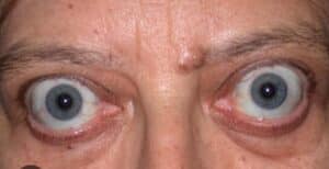
A 35-year-old woman presents for dental treatment with bilateral exophthalmos, weight loss, heat intolerance, and tachycardia. Which of the following systemic conditions is most commonly associated with these clinical findings?
Correct
ANSWER
Graves diseaseOTHER OPTIONS
● Cleidocranial dysostosis – Characterized by clavicular hypoplasia, delayed eruption of permanent teeth, and multiple supernumerary teeth; exophthalmos is not a typical feature.
● Peutz-Jeghers syndrome – Characterized by mucocutaneous pigmentation and gastrointestinal hamartomatous polyps rather than exophthalmos.
● Down syndrome – Common features include intellectual disability, hypotonia, and characteristic craniofacial features, but exophthalmos is not a characteristic finding.SYNOPSIS
● Graves disease is an autoimmune disorder causing hyperthyroidism and is the most common cause of exophthalmos (thyroid eye disease).
● Clinical manifestations include exophthalmos, diffuse goiter, tachycardia, heat intolerance, weight loss, and tremors.
● Dental management should minimize stress, avoid excessive use of vasoconstrictors in uncontrolled hyperthyroidism, and postpone elective treatment in untreated or poorly controlled patients.REFERENCE
Little and Falace’s Dental Management of the Medically Compromised Patient – 10th EditionIncorrect
ANSWER
Graves diseaseOTHER OPTIONS
● Cleidocranial dysostosis – Characterized by clavicular hypoplasia, delayed eruption of permanent teeth, and multiple supernumerary teeth; exophthalmos is not a typical feature.
● Peutz-Jeghers syndrome – Characterized by mucocutaneous pigmentation and gastrointestinal hamartomatous polyps rather than exophthalmos.
● Down syndrome – Common features include intellectual disability, hypotonia, and characteristic craniofacial features, but exophthalmos is not a characteristic finding.SYNOPSIS
● Graves disease is an autoimmune disorder causing hyperthyroidism and is the most common cause of exophthalmos (thyroid eye disease).
● Clinical manifestations include exophthalmos, diffuse goiter, tachycardia, heat intolerance, weight loss, and tremors.
● Dental management should minimize stress, avoid excessive use of vasoconstrictors in uncontrolled hyperthyroidism, and postpone elective treatment in untreated or poorly controlled patients.REFERENCE
Little and Falace’s Dental Management of the Medically Compromised Patient – 10th Edition -
Question 60 of 150
60. Question
A 52-year-old patient undergoing chemotherapy for a solid malignancy presents for a Class II composite restoration. Recent laboratory investigations reveal a normal complete blood count, including an adequate absolute neutrophil count and platelet count. The patient’s oncologist has confirmed that there are no contraindications to routine dental care. What is the most appropriate management?
Correct
ANSWER
Proceed with the planned restorationOTHER OPTIONS
● Defer dental treatment until completion of chemotherapy – Elective restorative treatment may be performed when hematologic parameters are within acceptable limits and the patient is medically stable.
● Prescribe prophylactic antibiotics before treatment – Routine antibiotic prophylaxis is not indicated solely because a patient is receiving chemotherapy if the neutrophil count is normal and there is no other indication.
● Prescribe analgesics and refer the patient back to the oncologist – Referral without treatment is unnecessary when the patient is medically stable and laboratory values are within normal limits.SYNOPSIS
● Routine restorative dental treatment can be safely performed in patients undergoing chemotherapy when the complete blood count, particularly the absolute neutrophil count and platelet count, is within acceptable limits.
● Consultation with the oncologist is recommended to determine the optimal timing of treatment and to confirm medical stability.
● Antibiotic prophylaxis is reserved for patients with significant neutropenia or other specific medical indications and is not routinely required.REFERENCE
Little and Falace’s Dental Management of the Medically Compromised Patient – 10th EditionIncorrect
ANSWER
Proceed with the planned restorationOTHER OPTIONS
● Defer dental treatment until completion of chemotherapy – Elective restorative treatment may be performed when hematologic parameters are within acceptable limits and the patient is medically stable.
● Prescribe prophylactic antibiotics before treatment – Routine antibiotic prophylaxis is not indicated solely because a patient is receiving chemotherapy if the neutrophil count is normal and there is no other indication.
● Prescribe analgesics and refer the patient back to the oncologist – Referral without treatment is unnecessary when the patient is medically stable and laboratory values are within normal limits.SYNOPSIS
● Routine restorative dental treatment can be safely performed in patients undergoing chemotherapy when the complete blood count, particularly the absolute neutrophil count and platelet count, is within acceptable limits.
● Consultation with the oncologist is recommended to determine the optimal timing of treatment and to confirm medical stability.
● Antibiotic prophylaxis is reserved for patients with significant neutropenia or other specific medical indications and is not routinely required.REFERENCE
Little and Falace’s Dental Management of the Medically Compromised Patient – 10th Edition -
Question 61 of 150
61. Question
A 35-year-old patient with human immunodeficiency virus (HIV) infection presents for dental treatment. A recent laboratory report shows a markedly reduced CD4+ T-lymphocyte count. According to the diagnostic criteria for acquired immunodeficiency syndrome (AIDS), a CD4+ cell count below which value is indicative of severe immunosuppression?
Correct
ANSWER
200OTHER OPTIONS
Not applicableSYNOPSIS
● The normal CD4+ T-lymphocyte count ranges from approximately 500 to 1,500 cells/mm³.
● A CD4+ count <200 cells/mm³ or the presence of an AIDS-defining illness is the accepted criterion for the diagnosis of AIDS.
● Patients with low CD4+ counts are at increased risk of opportunistic infections, and dental treatment should be planned after assessing immune status and overall medical condition.REFERENCE
Little and Falace's Dental Management of the Medically Compromised Patient - 10th EditionIncorrect
ANSWER
200OTHER OPTIONS
Not applicableSYNOPSIS
● The normal CD4+ T-lymphocyte count ranges from approximately 500 to 1,500 cells/mm³.
● A CD4+ count <200 cells/mm³ or the presence of an AIDS-defining illness is the accepted criterion for the diagnosis of AIDS.
● Patients with low CD4+ counts are at increased risk of opportunistic infections, and dental treatment should be planned after assessing immune status and overall medical condition.REFERENCE
Little and Falace's Dental Management of the Medically Compromised Patient - 10th Edition -
Question 62 of 150
62. Question
A 65-year-old patient with a long history of smoking is diagnosed with emphysema, characterized by permanent enlargement and destruction of the alveolar air spaces. The patient complains of persistent breathlessness, especially during exertion. What is the medical term for this symptom?
Correct
ANSWER
DyspneaOTHER OPTIONS
● Chronic cough – A common symptom of chronic obstructive pulmonary disease (COPD), but it is not synonymous with breathlessness.
● Cyanosis – Refers to bluish discoloration of the skin and mucous membranes due to inadequate oxygenation, not breathlessness.
● Dysphagia – Refers to difficulty in swallowing and is unrelated to emphysema-induced respiratory symptoms.SYNOPSIS
● Emphysema is a form of chronic obstructive pulmonary disease (COPD) characterized by destruction of alveolar walls and enlargement of distal air spaces.
● Dyspnea (breathlessness) is the hallmark clinical feature of emphysema and typically worsens with exertion as lung function declines.
● Dental management should include short appointments, stress reduction, upright chair positioning, and avoidance of respiratory depressants in patients with severe emphysema.REFERENCE
Little and Falace’s Dental Management of the Medically Compromised Patient – 10th EditionIncorrect
ANSWER
DyspneaOTHER OPTIONS
● Chronic cough – A common symptom of chronic obstructive pulmonary disease (COPD), but it is not synonymous with breathlessness.
● Cyanosis – Refers to bluish discoloration of the skin and mucous membranes due to inadequate oxygenation, not breathlessness.
● Dysphagia – Refers to difficulty in swallowing and is unrelated to emphysema-induced respiratory symptoms.SYNOPSIS
● Emphysema is a form of chronic obstructive pulmonary disease (COPD) characterized by destruction of alveolar walls and enlargement of distal air spaces.
● Dyspnea (breathlessness) is the hallmark clinical feature of emphysema and typically worsens with exertion as lung function declines.
● Dental management should include short appointments, stress reduction, upright chair positioning, and avoidance of respiratory depressants in patients with severe emphysema.REFERENCE
Little and Falace’s Dental Management of the Medically Compromised Patient – 10th Edition -
Question 63 of 150
63. Question
Failure of fusion of the palatal shelves during embryonic development results in cleft palate. During which period of intrauterine life does this developmental defect most commonly occur?
Correct
ANSWER
9th to 11th weekOTHER OPTIONS
● 6th week – This period is primarily associated with the development and fusion of the upper lip.
● 6th to 8th week – Failure of fusion during this period is more commonly associated with cleft lip rather than cleft palate.
● 10th week – Palatal fusion occurs over a range of weeks rather than at a single point in time.SYNOPSIS
● The secondary palate is formed by fusion of the palatal shelves derived from the maxillary processes.
● Fusion of the palatal shelves normally occurs between the 9th and 11th weeks of intrauterine life.
● Failure of this fusion results in cleft palate, which may occur alone or in association with cleft lip.REFERENCE
Langman’s Medical Embryology – 15th Edition.Incorrect
ANSWER
9th to 11th weekOTHER OPTIONS
● 6th week – This period is primarily associated with the development and fusion of the upper lip.
● 6th to 8th week – Failure of fusion during this period is more commonly associated with cleft lip rather than cleft palate.
● 10th week – Palatal fusion occurs over a range of weeks rather than at a single point in time.SYNOPSIS
● The secondary palate is formed by fusion of the palatal shelves derived from the maxillary processes.
● Fusion of the palatal shelves normally occurs between the 9th and 11th weeks of intrauterine life.
● Failure of this fusion results in cleft palate, which may occur alone or in association with cleft lip.REFERENCE
Langman’s Medical Embryology – 15th Edition. -
Question 64 of 150
64. Question
A patient sustains a gunshot injury resulting in loss of the mandibular condyle. Which of the following grafts is most commonly used for reconstruction of the condyle?
Correct
ANSWER
Costochondral graftOTHER OPTIONS
● Endochondral graft – Endochondral ossification refers to a mechanism of bone formation and is not a specific graft type used for condylar reconstruction.
● Periosteal graft – Periosteal grafts lack the structural support required for condylar reconstruction.SYNOPSIS
● Costochondral grafts harvested from the rib are widely used for reconstruction of the mandibular condyle following trauma, ankylosis, tumor resection, or gunshot injuries.
● These grafts provide both bone and cartilage components, closely resembling the structure and function of the mandibular condyle.
● In growing patients, costochondral grafts possess growth potential and are considered the gold standard for autogenous condylar reconstruction.REFERENCE
Peterson’s Principles of Oral and Maxillofacial Surgery – 7th Edition.Incorrect
ANSWER
Costochondral graftOTHER OPTIONS
● Endochondral graft – Endochondral ossification refers to a mechanism of bone formation and is not a specific graft type used for condylar reconstruction.
● Periosteal graft – Periosteal grafts lack the structural support required for condylar reconstruction.SYNOPSIS
● Costochondral grafts harvested from the rib are widely used for reconstruction of the mandibular condyle following trauma, ankylosis, tumor resection, or gunshot injuries.
● These grafts provide both bone and cartilage components, closely resembling the structure and function of the mandibular condyle.
● In growing patients, costochondral grafts possess growth potential and are considered the gold standard for autogenous condylar reconstruction.REFERENCE
Peterson’s Principles of Oral and Maxillofacial Surgery – 7th Edition. -
Question 65 of 150
65. Question
A patient undergoes oral surgery and returns for follow-up. The clinician explains that adequate revascularization of the wound is essential for successful healing. Approximately how long does it take for vascularity to become established during normal wound healing?
Correct
ANSWER
7–14 daysOTHER OPTIONS
● Not applicableSYNOPSIS
● Wound healing proceeds through inflammatory, proliferative, and remodeling phases.
● During the proliferative phase, angiogenesis and granulation tissue formation result in re-establishment of vascularity.
● Functional vascularization of the wound is generally achieved within 7–14 days, which is critical for tissue repair and resistance to infection.REFERENCE
Peterson’s Principles of Oral and Maxillofacial Surgery – 7th Edition.Incorrect
ANSWER
7–14 daysOTHER OPTIONS
● Not applicableSYNOPSIS
● Wound healing proceeds through inflammatory, proliferative, and remodeling phases.
● During the proliferative phase, angiogenesis and granulation tissue formation result in re-establishment of vascularity.
● Functional vascularization of the wound is generally achieved within 7–14 days, which is critical for tissue repair and resistance to infection.REFERENCE
Peterson’s Principles of Oral and Maxillofacial Surgery – 7th Edition. -
Question 66 of 150
66. Question
How is bone swaging defined?
Correct
ANSWER
Pushing bone into contact with the root surface without fracturing the bone at its baseOTHER OPTIONS
● Pushing bone into contact with the root surface with fracturing the bone at its base – Bone swaging preserves the base of the bone and maintains its blood supply.
● Removal of bone from the root surface during periodontal surgery – This describes osseous resective procedures rather than bone swaging.
● A technique used for saucerization of bone – Saucerization involves reshaping bony architecture and is distinct from bone swaging.SYNOPSIS
● Bone swaging is a regenerative osseous procedure in which a section of bone adjacent to an intrabony defect is repositioned against the root surface.
● The bone is displaced without fracturing its base, thereby preserving vascularity and promoting bone regeneration.
● Bone swaging is indicated when sufficient thickness of interproximal bone is present adjacent to a vertical defect.REFERENCE
Carranza’s Clinical Periodontology – 14th Edition.Incorrect
ANSWER
Pushing bone into contact with the root surface without fracturing the bone at its baseOTHER OPTIONS
● Pushing bone into contact with the root surface with fracturing the bone at its base – Bone swaging preserves the base of the bone and maintains its blood supply.
● Removal of bone from the root surface during periodontal surgery – This describes osseous resective procedures rather than bone swaging.
● A technique used for saucerization of bone – Saucerization involves reshaping bony architecture and is distinct from bone swaging.SYNOPSIS
● Bone swaging is a regenerative osseous procedure in which a section of bone adjacent to an intrabony defect is repositioned against the root surface.
● The bone is displaced without fracturing its base, thereby preserving vascularity and promoting bone regeneration.
● Bone swaging is indicated when sufficient thickness of interproximal bone is present adjacent to a vertical defect.REFERENCE
Carranza’s Clinical Periodontology – 14th Edition. -
Question 67 of 150
67. Question
Which of the following measures is most effective in reducing the occurrence of osteoradionecrosis in irradiated jaws?
Correct
ANSWER
Extraction with hyperbaric oxygen therapyOTHER OPTIONS
● Endodontic treatment and periodontal curettage – Conservative dental treatment is preferred to avoid extractions but does not specifically reduce the risk once extraction is required.
● Enucleation – This is a surgical procedure for cyst removal and has no role in preventing osteoradionecrosis.
● Marsupialization – This technique is used for decompression of cysts and is unrelated to the prevention of osteoradionecrosis.SYNOPSIS
● Osteoradionecrosis is a serious complication of radiation therapy characterized by hypovascular, hypocellular, and hypoxic bone.
● When dental extraction is unavoidable in irradiated patients, the risk of osteoradionecrosis can be reduced by using hyperbaric oxygen therapy (HBOT) in conjunction with atraumatic extraction techniques.
● Hyperbaric oxygen therapy enhances angiogenesis and tissue oxygenation, thereby promoting wound healing and decreasing the incidence of osteoradionecrosis.REFERENCE
Peterson’s Principles of Oral and Maxillofacial Surgery – 7th Edition.Incorrect
ANSWER
Extraction with hyperbaric oxygen therapyOTHER OPTIONS
● Endodontic treatment and periodontal curettage – Conservative dental treatment is preferred to avoid extractions but does not specifically reduce the risk once extraction is required.
● Enucleation – This is a surgical procedure for cyst removal and has no role in preventing osteoradionecrosis.
● Marsupialization – This technique is used for decompression of cysts and is unrelated to the prevention of osteoradionecrosis.SYNOPSIS
● Osteoradionecrosis is a serious complication of radiation therapy characterized by hypovascular, hypocellular, and hypoxic bone.
● When dental extraction is unavoidable in irradiated patients, the risk of osteoradionecrosis can be reduced by using hyperbaric oxygen therapy (HBOT) in conjunction with atraumatic extraction techniques.
● Hyperbaric oxygen therapy enhances angiogenesis and tissue oxygenation, thereby promoting wound healing and decreasing the incidence of osteoradionecrosis.REFERENCE
Peterson’s Principles of Oral and Maxillofacial Surgery – 7th Edition. -
Question 68 of 150
68. Question
From where does the bleeding occurs mainly in LEFORT 1 fracture?
Correct
ANSWER
Maxillary arteryOTHER OPTIONS
● Infraorbital artery – More commonly involved in Le Fort II fractures due to infraorbital rim involvement.
● Mandibular artery – Not a recognized major artery associated with Le Fort I fractures.
● Facial artery – Supplies superficial facial structures and is not the principal source of bleeding in Le Fort I fractures.SYNOPSIS
● Le Fort I fracture is a horizontal fracture of the maxilla extending through the lateral wall of the maxillary sinus and pterygomaxillary region.
● Significant hemorrhage in Le Fort I fractures commonly originates from branches of the maxillary artery, especially the descending palatine branch.
● Injury to these vessels can lead to severe palatal and nasal bleeding following maxillary fractures.REFERENCE
Peterson’s Principles of Oral and Maxillofacial Surgery – 7th EditionIncorrect
ANSWER
Maxillary arteryOTHER OPTIONS
● Infraorbital artery – More commonly involved in Le Fort II fractures due to infraorbital rim involvement.
● Mandibular artery – Not a recognized major artery associated with Le Fort I fractures.
● Facial artery – Supplies superficial facial structures and is not the principal source of bleeding in Le Fort I fractures.SYNOPSIS
● Le Fort I fracture is a horizontal fracture of the maxilla extending through the lateral wall of the maxillary sinus and pterygomaxillary region.
● Significant hemorrhage in Le Fort I fractures commonly originates from branches of the maxillary artery, especially the descending palatine branch.
● Injury to these vessels can lead to severe palatal and nasal bleeding following maxillary fractures.REFERENCE
Peterson’s Principles of Oral and Maxillofacial Surgery – 7th Edition -
Question 69 of 150
69. Question
A 50-year-old male patient presents with swelling and severe pain in the lower left jaw. He reports the pain started three days ago and has progressively worsened. Clinical examination reveals localized swelling and tenderness in the region of the affected tooth. The patient has a history of Type 2 diabetes, and his random blood sugar (RBS) reading at presentation is 20 mmol/L (360 mg/dL). He requests an immediate extraction of the tooth. What is the most appropriate management in this case?
Correct
ANSWER
Prescribe analgesics and antibioticsOTHER OPTIONS
• Prescribe analgesics – While analgesics will provide temporary pain relief, they do not address the infection or the elevated blood sugar levels, making this option insufficient.
• Postpone treatment – Immediate treatment of the infection is necessary as uncontrolled diabetes can worsen the condition. Delaying management without addressing the infection is not appropriate.
• Prescribe antibiotics – Antibiotics alone are insufficient for pain relief and managing symptoms, as they do not address the patient’s immediate discomfort.SYNOPSIS
• Patients with diabetes and infections require cautious management.
• The elevated RBS indicates poor glycemic control, which can impair healing and exacerbate the infection.
• Immediate extraction is contraindicated until the infection is managed and blood sugar levels are better controlled.
• Prescribing a combination of antibiotics to manage the infection and analgesics for pain relief is appropriate.
• Extraction can be performed after the infection subsides, and glycemic levels are stabilized.
• Coordination with the patient’s physician for blood sugar control is crucial.REFERENCE
Malamed, S. F. (2019). Handbook of Local Anesthesia (7th ed.). Elsevier.
Greenberg, M. S., Glick, M., & Ship, J. A. (2019). Burket’s Oral Medicine (13th ed.). People’s Medical Publishing House.Incorrect
ANSWER
Prescribe analgesics and antibioticsOTHER OPTIONS
• Prescribe analgesics – While analgesics will provide temporary pain relief, they do not address the infection or the elevated blood sugar levels, making this option insufficient.
• Postpone treatment – Immediate treatment of the infection is necessary as uncontrolled diabetes can worsen the condition. Delaying management without addressing the infection is not appropriate.
• Prescribe antibiotics – Antibiotics alone are insufficient for pain relief and managing symptoms, as they do not address the patient’s immediate discomfort.SYNOPSIS
• Patients with diabetes and infections require cautious management.
• The elevated RBS indicates poor glycemic control, which can impair healing and exacerbate the infection.
• Immediate extraction is contraindicated until the infection is managed and blood sugar levels are better controlled.
• Prescribing a combination of antibiotics to manage the infection and analgesics for pain relief is appropriate.
• Extraction can be performed after the infection subsides, and glycemic levels are stabilized.
• Coordination with the patient’s physician for blood sugar control is crucial.REFERENCE
Malamed, S. F. (2019). Handbook of Local Anesthesia (7th ed.). Elsevier.
Greenberg, M. S., Glick, M., & Ship, J. A. (2019). Burket’s Oral Medicine (13th ed.). People’s Medical Publishing House. -
Question 70 of 150
70. Question
A 15-year-old patient undergoes apexification treatment for an immature permanent tooth with a necrotic pulp. Non-setting calcium hydroxide is placed in the root canal to induce hard tissue formation before applying Mineral Trioxide Aggregate (MTA). What is the recommended time interval for replacing the non-setting calcium hydroxide dressing before proceeding with MTA placement?
Correct
ANSWER
2 weeksOTHER OPTIONS
• 6 months – Leaving calcium hydroxide in the canal for this long may weaken dentin and increase the risk of fracture.SYNOPSIS
• Apexification using Ca(OH)₂ as the main barrier-inducing material – Replaced every 3 months until barrier forms (6–24 months total)
• Short-term Ca(OH)₂ before MTA apical plug 1–2 weeks onlyREFERENCE
Hargreaves, K. M., & Berman, L. H. (2016). Cohen’s Pathways of the Pulp (11th ed.). Elsevier.Incorrect
ANSWER
2 weeksOTHER OPTIONS
• 6 months – Leaving calcium hydroxide in the canal for this long may weaken dentin and increase the risk of fracture.SYNOPSIS
• Apexification using Ca(OH)₂ as the main barrier-inducing material – Replaced every 3 months until barrier forms (6–24 months total)
• Short-term Ca(OH)₂ before MTA apical plug 1–2 weeks onlyREFERENCE
Hargreaves, K. M., & Berman, L. H. (2016). Cohen’s Pathways of the Pulp (11th ed.). Elsevier. -
Question 71 of 150
71. Question
45-year-old patient presents for a routine dental check-up. Upon examination, the dental hygienist notes that the patient has approximately 30% plaque accumulation on their teeth and a clinical attachment loss (CAL) of 4-5 mm in some areas. The patient has been educated on oral hygiene practices in previous visits but admits to not consistently following the recommended oral care routine at home. The dentist decides to assess the patient’s stage of change to determine the most appropriate approach for treatment and education. Based on the patient’s current behavior and awareness, which stage of change is the patient most likely in?
Correct
ANSWER
ContemplationOTHER OPTIONS
• Refer SynopsisSYNOPSIS
• The stage of change model, or Transtheoretical Model, consists of several stages – precontemplation, contemplation, preparation, action, and maintenance.
• In the contemplation stage, the patient is aware of their problem (in this case, plaque accumulation and clinical attachment loss) and is thinking about making a change but has not yet taken significant action.
• The patient admits to inconsistent oral hygiene but is likely aware of the need to improve.
• This stage typically involves ambivalence-recognizing the need for change but not fully committing to it.
• Precontemplation occurs when the patient is not yet aware of or is in denial about their oral health issues.
• Relapse would apply if the patient had previously made significant progress in improving their oral hygiene but had fallen back into poor habits.
• Maintenance occurs after the patient has made changes and is working to sustain the new behaviors.REFERENCE
Oral Health Promotion and Disease Prevention by David W. Jones.Incorrect
ANSWER
ContemplationOTHER OPTIONS
• Refer SynopsisSYNOPSIS
• The stage of change model, or Transtheoretical Model, consists of several stages – precontemplation, contemplation, preparation, action, and maintenance.
• In the contemplation stage, the patient is aware of their problem (in this case, plaque accumulation and clinical attachment loss) and is thinking about making a change but has not yet taken significant action.
• The patient admits to inconsistent oral hygiene but is likely aware of the need to improve.
• This stage typically involves ambivalence-recognizing the need for change but not fully committing to it.
• Precontemplation occurs when the patient is not yet aware of or is in denial about their oral health issues.
• Relapse would apply if the patient had previously made significant progress in improving their oral hygiene but had fallen back into poor habits.
• Maintenance occurs after the patient has made changes and is working to sustain the new behaviors.REFERENCE
Oral Health Promotion and Disease Prevention by David W. Jones. -
Question 72 of 150
72. Question
65-year-old patient with a history of atrial fibrillation visits the dental clinic for the extraction of a lower molar. The patient is on a daily morning dose of an oral anticoagulant (e.g., warfarin or a direct oral anticoagulant like apixaban or rivaroxaban) to prevent thromboembolic events. The dentist needs to decide whether the medication should be stopped or adjusted before the procedure to minimize the risk of bleeding complications while maintaining thromboembolic protection. What is the appropriate management of the patient’s anticoagulant medication before the dental extraction?
Correct
ANSWER
No need to stop the anticoagulant.OTHER OPTIONS
• Not applicableSYNOPSIS
• For most routine dental extractions, there is no need to stop anticoagulant medication in patients with atrial fibrillation.
• Studies have shown that the risk of significant bleeding is minimal when the extraction is performed under local measures, such as using hemostatic agents (e.g., collagen sponges, sutures) and applying pressure post-operatively.
• Stopping anticoagulants increases the risk of thromboembolic events, which can be life-threatening.
• The dentist should consult the patient’s physician if there is any uncertainty, but routine anticoagulant therapy should generally be continued.REFERENCE
Guidelines on the Management of Anticoagulant Therapy in Dental Procedures by the American College of Cardiology.Incorrect
ANSWER
No need to stop the anticoagulant.OTHER OPTIONS
• Not applicableSYNOPSIS
• For most routine dental extractions, there is no need to stop anticoagulant medication in patients with atrial fibrillation.
• Studies have shown that the risk of significant bleeding is minimal when the extraction is performed under local measures, such as using hemostatic agents (e.g., collagen sponges, sutures) and applying pressure post-operatively.
• Stopping anticoagulants increases the risk of thromboembolic events, which can be life-threatening.
• The dentist should consult the patient’s physician if there is any uncertainty, but routine anticoagulant therapy should generally be continued.REFERENCE
Guidelines on the Management of Anticoagulant Therapy in Dental Procedures by the American College of Cardiology. -
Question 73 of 150
73. Question
Which of the following diseases is commonly caused by the Epstein-Barr Virus (EBV)?
Correct
ANSWER
Infectious mononucleosisOTHER OPTIONS
• Oral candidiasis – This is caused by Candida albicans, a fungal infection, not EBV.
• Herpangina – This is caused by Coxsackievirus, not EBV.
• Kaposi sarcoma – This is associated with Human Herpesvirus 8 (HHV-8), not EBV.SYNOPSIS
• The Epstein-Barr Virus (EBV) is a member of the herpesvirus family and is known to cause several diseases.
• The most common condition caused by EBV is infectious mononucleosis, which is characterized by fever, lymphadenopathy, pharyngitis, and fatigue.
• EBV also has a strong association with other conditions, including
– Burkitt’s lymphoma (a type of non-Hodgkin lymphoma).
– Nasopharyngeal carcinoma (common in certain populations).
– Hodgkin’s lymphoma (certain subtypes).
– Oral hairy leukoplakia (seen in immunocompromised patients, such as those with HIV).REFERENCE
Jawetz, Melnick & Adelberg’s Medical Microbiology, 28th Edition.
Epstein-Barr Virus and Human Disease in The Lancet by Cohen, J. I. (2021).Incorrect
ANSWER
Infectious mononucleosisOTHER OPTIONS
• Oral candidiasis – This is caused by Candida albicans, a fungal infection, not EBV.
• Herpangina – This is caused by Coxsackievirus, not EBV.
• Kaposi sarcoma – This is associated with Human Herpesvirus 8 (HHV-8), not EBV.SYNOPSIS
• The Epstein-Barr Virus (EBV) is a member of the herpesvirus family and is known to cause several diseases.
• The most common condition caused by EBV is infectious mononucleosis, which is characterized by fever, lymphadenopathy, pharyngitis, and fatigue.
• EBV also has a strong association with other conditions, including
– Burkitt’s lymphoma (a type of non-Hodgkin lymphoma).
– Nasopharyngeal carcinoma (common in certain populations).
– Hodgkin’s lymphoma (certain subtypes).
– Oral hairy leukoplakia (seen in immunocompromised patients, such as those with HIV).REFERENCE
Jawetz, Melnick & Adelberg’s Medical Microbiology, 28th Edition.
Epstein-Barr Virus and Human Disease in The Lancet by Cohen, J. I. (2021). -
Question 74 of 150
74. Question
30-year-old patient with Angle’s Class II malocclusion presents with moderate crowding in the lower arch and a deep overbite.In a 30-year-old patient with Angle’s Class II malocclusion, which of the following would be the most appropriate extraction choice to address crowding and achieve ideal occlusion?
Correct
ANSWER
Extraction of maxillary first premolarsOTHER OPTIONS
• Extraction of mandibular first premolars – This is less commonly done in Class II cases as it may worsen the skeletal imbalance and is more appropriate for Class I cases or severe crowding in the lower arch.
• Extraction of maxillary third molars – Third molar extractions are typically not performed to treat crowding or malocclusion unless there are specific concerns regarding impaction or pathology, and they do not directly address the occlusal issues seen in Class II malocclusion.
• No extraction necessary – In some cases of mild Class II malocclusion, no extraction may be needed, but in a patient with moderate crowding and deep overbite, extractions are often required to achieve ideal space and occlusion.SYNOPSIS
• In Angle’s Class II malocclusion, especially with moderate crowding, the typical approach often involves extracting maxillary first premolars to create space for aligning the anterior teeth and improving the overbite.
• This helps in correcting the overjet, aligning the upper and lower teeth, and improving the occlusion.
• The upper arch typically benefits from the extraction of first premolars in Class II cases because this moves the upper teeth back to reduce the overjet and helps achieve better facial balance.REFERENCE
Contemporary Orthodontics by William R. Proffit, Henry W. Fields Jr., and David M. Sarver.
Principles of Orthodontics – Extraction or Non-extraction Treatment? – Journal of Orthodontics and Dentofacial Orthopedics by Bishara, S. E.Incorrect
ANSWER
Extraction of maxillary first premolarsOTHER OPTIONS
• Extraction of mandibular first premolars – This is less commonly done in Class II cases as it may worsen the skeletal imbalance and is more appropriate for Class I cases or severe crowding in the lower arch.
• Extraction of maxillary third molars – Third molar extractions are typically not performed to treat crowding or malocclusion unless there are specific concerns regarding impaction or pathology, and they do not directly address the occlusal issues seen in Class II malocclusion.
• No extraction necessary – In some cases of mild Class II malocclusion, no extraction may be needed, but in a patient with moderate crowding and deep overbite, extractions are often required to achieve ideal space and occlusion.SYNOPSIS
• In Angle’s Class II malocclusion, especially with moderate crowding, the typical approach often involves extracting maxillary first premolars to create space for aligning the anterior teeth and improving the overbite.
• This helps in correcting the overjet, aligning the upper and lower teeth, and improving the occlusion.
• The upper arch typically benefits from the extraction of first premolars in Class II cases because this moves the upper teeth back to reduce the overjet and helps achieve better facial balance.REFERENCE
Contemporary Orthodontics by William R. Proffit, Henry W. Fields Jr., and David M. Sarver.
Principles of Orthodontics – Extraction or Non-extraction Treatment? – Journal of Orthodontics and Dentofacial Orthopedics by Bishara, S. E. -
Question 75 of 150
75. Question
9-year-old child presents to the clinic following a traumatic dental injury. Clinical examination reveals that the maxillary right central incisor has been intruded into the alveolar bone by approximately 7 mm. Radiographic findings confirm no root fracture.How would this intrusion injury be classified based on severity?
Correct
ANSWER
SevereOTHER OPTIONS
• Refer SynopsisSYNOPSIS
• Traumatic dental injuries such as intrusion are classified based on the degree of intrusion
• Mild – Intrusion of less than 3 mm in primary or permanent teeth.
• Moderate – Intrusion of 3–6 mm in permanent teeth.
• Severe – Intrusion of greater than 6 mm in permanent teeth.
• Very Severe – Rarely used clinically, may describe complete displacement into the alveolar bone.
• Since the intrusion depth in this scenario is 7 mm, it falls into the severe category.REFERENCE
Chapter 8 – Intrusive Luxation -Traumatic Dental Injuries – A Manual, 3rd Edition. Andreasen JO, Andreasen FM, Andersson L.Incorrect
ANSWER
SevereOTHER OPTIONS
• Refer SynopsisSYNOPSIS
• Traumatic dental injuries such as intrusion are classified based on the degree of intrusion
• Mild – Intrusion of less than 3 mm in primary or permanent teeth.
• Moderate – Intrusion of 3–6 mm in permanent teeth.
• Severe – Intrusion of greater than 6 mm in permanent teeth.
• Very Severe – Rarely used clinically, may describe complete displacement into the alveolar bone.
• Since the intrusion depth in this scenario is 7 mm, it falls into the severe category.REFERENCE
Chapter 8 – Intrusive Luxation -Traumatic Dental Injuries – A Manual, 3rd Edition. Andreasen JO, Andreasen FM, Andersson L. -
Question 76 of 150
76. Question
25-year-old patient with sickle cell anemia visits the dental clinic for extraction of a grossly decayed mandibular molar. The patient reports frequent vaso-occlusive crises requiring hospitalization but is currently stable. Vital signs are within normal limits, and no acute symptoms are present during the consultation. What is the ASA (American Society of Anesthesiologists) classification for this patient?
Correct
ANSWER
ASA IIIOTHER OPTIONS
• Refer SynopsisSYNOPSIS
• The ASA classification system is used to determine the physical status of a patient and assess their anesthetic risk. It ranges from ASA I (healthy patient) to ASA VI (brain-dead patient).
– ASA I – A healthy patient with no systemic disease.
– ASA II – A patient with mild systemic disease that does not limit daily activities (e.g., controlled hypertension, mild asthma).
– ASA III – A patient with severe systemic disease that limits daily activity but is not incapacitating (e.g., poorly controlled diabetes, sickle cell anemia with frequent crises).
– ASA IV – A patient with severe systemic disease that is a constant threat to life (e.g., unstable angina, recent myocardial infarction).
• The patient has sickle cell anemia, a severe systemic condition.
• Frequent vaso-occlusive crises indicate the condition significantly impacts daily life.
• However, the patient is stable at the time of the consultation and not in acute crisis, so they do not fall into the ASA IV category.
• Thus, the patient is classified as ASA III.REFERENCE
Chapter 1 – Patient Assessment and Risk Management – Malamed SF. Handbook of Local Anesthesia, 7th Edition,Incorrect
ANSWER
ASA IIIOTHER OPTIONS
• Refer SynopsisSYNOPSIS
• The ASA classification system is used to determine the physical status of a patient and assess their anesthetic risk. It ranges from ASA I (healthy patient) to ASA VI (brain-dead patient).
– ASA I – A healthy patient with no systemic disease.
– ASA II – A patient with mild systemic disease that does not limit daily activities (e.g., controlled hypertension, mild asthma).
– ASA III – A patient with severe systemic disease that limits daily activity but is not incapacitating (e.g., poorly controlled diabetes, sickle cell anemia with frequent crises).
– ASA IV – A patient with severe systemic disease that is a constant threat to life (e.g., unstable angina, recent myocardial infarction).
• The patient has sickle cell anemia, a severe systemic condition.
• Frequent vaso-occlusive crises indicate the condition significantly impacts daily life.
• However, the patient is stable at the time of the consultation and not in acute crisis, so they do not fall into the ASA IV category.
• Thus, the patient is classified as ASA III.REFERENCE
Chapter 1 – Patient Assessment and Risk Management – Malamed SF. Handbook of Local Anesthesia, 7th Edition, -
Question 77 of 150
77. Question
35-year-old patient visits the dental clinic for a minor surgical procedure. The dentist plans to use nitrous oxide sedation for pain and anxiety management. The patient reports a history of chronic sinus infections, as well as recent treatment for pneumothorax 3 months ago. No other systemic conditions are reported. In which condition is nitrous oxide contraindicated for this patient?
Correct
ANSWER
Recent pneumothoraxOTHER OPTIONS
• Chronic sinus infections – Nitrous oxide can accumulate in sinuses, causing mild discomfort, but it is not strictly contraindicated.
• Mild anxiety – Nitrous oxide is commonly used for managing anxiety during dental procedures.
• History of dental phobia – Nitrous oxide is effective in calming phobic patients.SYNOPSIS
• Nitrous Oxide Contraindications:
• Nitrous oxide is generally safe for sedation but is contraindicated in conditions where gas expansion or pressure effects could be harmful due to its ability to diffuse into closed air spaces.
• Recent pneumothorax – Nitrous oxide diffuses into pleural spaces, significantly increasing pressure, which may exacerbate the condition or lead to recurrence. This makes pneumothorax a clear contraindication.
• In this case, the patient’s recent pneumothorax is the contraindication.REFERENCE
Chapter 7 – Nitrous Oxide-Oxygen Sedation – Malamed SF. Sedation – A Guide to Patient Management, 6th EditionIncorrect
ANSWER
Recent pneumothoraxOTHER OPTIONS
• Chronic sinus infections – Nitrous oxide can accumulate in sinuses, causing mild discomfort, but it is not strictly contraindicated.
• Mild anxiety – Nitrous oxide is commonly used for managing anxiety during dental procedures.
• History of dental phobia – Nitrous oxide is effective in calming phobic patients.SYNOPSIS
• Nitrous Oxide Contraindications:
• Nitrous oxide is generally safe for sedation but is contraindicated in conditions where gas expansion or pressure effects could be harmful due to its ability to diffuse into closed air spaces.
• Recent pneumothorax – Nitrous oxide diffuses into pleural spaces, significantly increasing pressure, which may exacerbate the condition or lead to recurrence. This makes pneumothorax a clear contraindication.
• In this case, the patient’s recent pneumothorax is the contraindication.REFERENCE
Chapter 7 – Nitrous Oxide-Oxygen Sedation – Malamed SF. Sedation – A Guide to Patient Management, 6th Edition -
Question 78 of 150
78. Question
65-year-old patient presents with discomfort while using a complete denture. Upon examination, the oral cavity shows significant bone resorption resulting in a flat alveolar ridge. The patient complains of repeated sore spots and instability of the denture. The dentist discusses the need for frequent tissue conditioning to improve comfort and fit. Which type of denture most commonly requires frequent tissue conditioning?
Correct
ANSWER
Denture on flat ridgeOTHER OPTIONS
• Denture on Tori Cases – Tori (bony outgrowths) can complicate denture fabrication and fitting, but once relieved properly during denture fabrication, tissue conditioning is less frequently required.
• Denture on Ridge with Sharp Bone – Sharp bony ridges can cause tissue trauma, but pre-prosthetic surgery to smoothen the ridge is often recommended before denture fabrication.SYNOPSIS
• A flat ridge results from extensive alveolar bone resorption, offering minimal support, retention, and stability for the denture.
• Due to poor ridge morphology, the denture tends to move, causing trauma to the soft tissues and necessitating frequent tissue conditioning to distribute pressure evenly and alleviate sore spots.REFERENCE
Chapter 5 Management of the Atrophic Ridge – Zarb GA, Bolender CL, Eckert SE. Prosthodontic Treatment for Edentulous Patients, 13th EditionIncorrect
ANSWER
Denture on flat ridgeOTHER OPTIONS
• Denture on Tori Cases – Tori (bony outgrowths) can complicate denture fabrication and fitting, but once relieved properly during denture fabrication, tissue conditioning is less frequently required.
• Denture on Ridge with Sharp Bone – Sharp bony ridges can cause tissue trauma, but pre-prosthetic surgery to smoothen the ridge is often recommended before denture fabrication.SYNOPSIS
• A flat ridge results from extensive alveolar bone resorption, offering minimal support, retention, and stability for the denture.
• Due to poor ridge morphology, the denture tends to move, causing trauma to the soft tissues and necessitating frequent tissue conditioning to distribute pressure evenly and alleviate sore spots.REFERENCE
Chapter 5 Management of the Atrophic Ridge – Zarb GA, Bolender CL, Eckert SE. Prosthodontic Treatment for Edentulous Patients, 13th Edition -
Question 79 of 150
79. Question
45-year-old female patient presents with complaints of persistent dry mouth and dry eyes. She reports difficulty swallowing food without water and has a history of fatigue and joint pain. Upon examination, there is evidence of dental caries and reduced salivary flow. The dentist suspects Sjögren syndrome and refers her for further diagnostic evaluation. Which of the following is a characteristic feature of Sjögren syndrome?
Correct
ANSWER
Dry mouth, dry eyes, and lymphocytic infiltration of salivary glandsOTHER OPTIONS
• Polyuria and polydipsia – Diabetes Mellitus
• Butterfly-shaped facial rash – SLE
• Severe generalized gingival hyperplasia – This is commonly associated with drug-induced changes (e.g., phenytoin, cyclosporine) or leukemia, not Sjögren syndrome.SYNOPSIS
• Sjögren syndrome is a chronic autoimmune disorder characterized by
– Xerostomia (Dry Mouth) -Caused by dysfunction or destruction of salivary glands due to lymphocytic infiltration.
– Keratoconjunctivitis Sicca (Dry Eyes) – Due to reduced tear secretion by the lacrimal glands.
– Systemic Features – Fatigue, joint pain, and other connective tissue diseases like rheumatoid arthritis or lupus.
– Histopathology – Lymphocytic infiltration of salivary glands is a hallmark feature.REFERENCE
Scully C. Oral and Maxillofacial Medicine – The Basis of Diagnosis and Treatment, 3rd Edition.Incorrect
ANSWER
Dry mouth, dry eyes, and lymphocytic infiltration of salivary glandsOTHER OPTIONS
• Polyuria and polydipsia – Diabetes Mellitus
• Butterfly-shaped facial rash – SLE
• Severe generalized gingival hyperplasia – This is commonly associated with drug-induced changes (e.g., phenytoin, cyclosporine) or leukemia, not Sjögren syndrome.SYNOPSIS
• Sjögren syndrome is a chronic autoimmune disorder characterized by
– Xerostomia (Dry Mouth) -Caused by dysfunction or destruction of salivary glands due to lymphocytic infiltration.
– Keratoconjunctivitis Sicca (Dry Eyes) – Due to reduced tear secretion by the lacrimal glands.
– Systemic Features – Fatigue, joint pain, and other connective tissue diseases like rheumatoid arthritis or lupus.
– Histopathology – Lymphocytic infiltration of salivary glands is a hallmark feature.REFERENCE
Scully C. Oral and Maxillofacial Medicine – The Basis of Diagnosis and Treatment, 3rd Edition. -
Question 80 of 150
80. Question
12-year-old patient presents for orthodontic evaluation. The patient has a moderate crowding of the upper and lower arch with a deep bite. The orthodontist is considering serial extraction as a potential treatment option to alleviate the crowding. However, the patient’s deep bite is concerning, and the orthodontist is unsure whether this procedure would be appropriate given the patient’s overall occlusal relationship. Which of the following is a contraindication for serial extraction?
Correct
ANSWER
Deep biteOTHER OPTIONS
• Not applicableSYNOPSIS
• Serial extraction is an orthodontic treatment protocol typically used in patients with moderate to severe crowding. It involves the planned extraction of specific primary and permanent teeth to guide the eruption of remaining teeth and create space for proper alignment.
• However, serial extraction is contraindicated in certain occlusal conditions due to the potential for exacerbating the existing malocclusion.
• Deep bite refers to excessive vertical overlap between the upper and lower incisors, which can lead to potential functional and aesthetic issues.
• Serial extractions can aggravate a deep bite, as extracting teeth to create space in a patient with a deep bite can further reduce the vertical dimension and may lead to an even more severe deep bite. T
• The procedure may lead to undesirable tooth movement, worsening the vertical overlap, and affecting occlusal stability in the long term.REFERENCE
Proffit WR, Fields HW, Sarver DM. Contemporary Orthodontics, 5th Edition, 2018.Incorrect
ANSWER
Deep biteOTHER OPTIONS
• Not applicableSYNOPSIS
• Serial extraction is an orthodontic treatment protocol typically used in patients with moderate to severe crowding. It involves the planned extraction of specific primary and permanent teeth to guide the eruption of remaining teeth and create space for proper alignment.
• However, serial extraction is contraindicated in certain occlusal conditions due to the potential for exacerbating the existing malocclusion.
• Deep bite refers to excessive vertical overlap between the upper and lower incisors, which can lead to potential functional and aesthetic issues.
• Serial extractions can aggravate a deep bite, as extracting teeth to create space in a patient with a deep bite can further reduce the vertical dimension and may lead to an even more severe deep bite. T
• The procedure may lead to undesirable tooth movement, worsening the vertical overlap, and affecting occlusal stability in the long term.REFERENCE
Proffit WR, Fields HW, Sarver DM. Contemporary Orthodontics, 5th Edition, 2018. -
Question 81 of 150
81. Question
30-year-old patient reports to the dental clinic with the complaint of improper occlusion after restorative dental procedures. The dentist plans to take an accurate bite registration to ensure proper alignment of the upper and lower arches. Which material is most suitable for bite restoration in this scenario?
Correct
ANSWER
ElastomerOTHER OPTIONS
• Hardwax – While it is an older material used for bite registrations, it can distort easily, especially under pressure or temperature changes, making it less reliable.
• Plaster of Paris – Though it provides accurate models, it is brittle and difficult to use directly for bite restoration.
• Zinc oxide-eugenol paste – Often used for its flow properties, but it is brittle once set, making it less durable for accurate bite restoration.SYNOPSIS
• Elastomeric materials, such as polyvinyl siloxane (PVS), are considered the best for bite registration due to their superior accuracy, dimensional stability, and ease of manipulation.
• These materials are soft enough to prevent distortion during occlusal contact but rigid enough to ensure precise recording of occlusal relationships.
• Elastomers set quickly, reducing the chance of patient movement affecting the accuracy.REFERENCE
Craig, R. G., & Powers, J. M. (2012). Restorative Dental Materials (13th ed.). Mosby.Incorrect
ANSWER
ElastomerOTHER OPTIONS
• Hardwax – While it is an older material used for bite registrations, it can distort easily, especially under pressure or temperature changes, making it less reliable.
• Plaster of Paris – Though it provides accurate models, it is brittle and difficult to use directly for bite restoration.
• Zinc oxide-eugenol paste – Often used for its flow properties, but it is brittle once set, making it less durable for accurate bite restoration.SYNOPSIS
• Elastomeric materials, such as polyvinyl siloxane (PVS), are considered the best for bite registration due to their superior accuracy, dimensional stability, and ease of manipulation.
• These materials are soft enough to prevent distortion during occlusal contact but rigid enough to ensure precise recording of occlusal relationships.
• Elastomers set quickly, reducing the chance of patient movement affecting the accuracy.REFERENCE
Craig, R. G., & Powers, J. M. (2012). Restorative Dental Materials (13th ed.). Mosby. -
Question 82 of 150
82. Question
What is the ideal angle between rest and minor connector?
Correct
ANSWER
Less than 90 degreesOTHER OPTIONS
• More than 90 degrees – An angle greater than 90 degrees fails to transmit occlusal forces along the supporting vertical axis of the abutment tooth. This also permits slippage of the prosthesis away from the abutment, which can result in orthodontic-like forces being applied to an inclined plane on the abutment, with possible tooth movement.SYNOPSIS
• The angle formed by the occlusal rest and the vertical minor connector from which it originates should be less than 90 degrees.
• Only in this way can the occlusal forces be directed along the long axis of the abutment tooth.REFERENCE
Chapter 6 Rests and Rest Seats. Pocket dentistry.Incorrect
ANSWER
Less than 90 degreesOTHER OPTIONS
• More than 90 degrees – An angle greater than 90 degrees fails to transmit occlusal forces along the supporting vertical axis of the abutment tooth. This also permits slippage of the prosthesis away from the abutment, which can result in orthodontic-like forces being applied to an inclined plane on the abutment, with possible tooth movement.SYNOPSIS
• The angle formed by the occlusal rest and the vertical minor connector from which it originates should be less than 90 degrees.
• Only in this way can the occlusal forces be directed along the long axis of the abutment tooth.REFERENCE
Chapter 6 Rests and Rest Seats. Pocket dentistry. -
Question 83 of 150
83. Question
What does bodily tooth movement mean in orthodontics?
Correct
ANSWER
Both A and COTHER OPTIONS
• Both the crown and root move in the same direction either labially or lingually the same amount of distance – Labiolingual direction is more of a tipping movementSYNOPSIS
• Bodily tooth movement in orthodontics refers to a type of tooth movement in which the entire tooth, including both the crown and the root, moves together as a unit in a horizontal direction.
• This is one of the most controlled and desirable forms of tooth movement during orthodontic treatment because the tooth shifts its position without significant tipping or rotation.REFERENCE
Biomechanics and esthetic strategies in clinical orthodontics-Nanda-Page 6.Incorrect
ANSWER
Both A and COTHER OPTIONS
• Both the crown and root move in the same direction either labially or lingually the same amount of distance – Labiolingual direction is more of a tipping movementSYNOPSIS
• Bodily tooth movement in orthodontics refers to a type of tooth movement in which the entire tooth, including both the crown and the root, moves together as a unit in a horizontal direction.
• This is one of the most controlled and desirable forms of tooth movement during orthodontic treatment because the tooth shifts its position without significant tipping or rotation.REFERENCE
Biomechanics and esthetic strategies in clinical orthodontics-Nanda-Page 6. -
Question 84 of 150
84. Question
What is the area of insertion in Gow-Gates injection technique?
Correct
ANSWER
Mucous membrane distal to second maxillary molar just below mesiopalatal cuspOTHER OPTIONS
• Mucous membrane taking pterygomandibular raphae and coronoid notch as landmarks – Inferior alveolar nerve block is given in mucous membrane taking pterygomandibular raphae and coronoid notch as landmarks.
• Adjacent to maxillary tuberosity at the height of mucogingival junction next to maxillary third molar – Vazirani akinosi is administered adjacent to maxillary tuberosity at the height of mucogingival junction next to maxillary third molar.
• Mucobuccal fold above maxillary second molar – Posterior superior alveolar nerve block is administered in mucobuccal fold above maxillary second molar.SYNOPSIS
• Mandibular nerve block or Gow Gates technique provides sensory anesthesia to virtually the entire distribution of mandibular nerve.
• It anesthetizes inferior alveolar, mental, incisive, lingual, mylohyoid, auriculotemporal and buccal nerves.
• Area of insertion is mucous membrane on mesial of mandibular ramus, mucosa distal to maxillary second molar by placement of needle tip just below the mesiopalatal cusp of maxillary second molar.
• Landmarks includes center of external auditory meatus and corner of mouth extraorally and intraorally mesiopalatal cusp of upper 2nd molar and soft tissues distal to 2nd molar.REFERENCE
Handbook of Local Anesthesia, Stanley F Malamed – 7th Edition Page No 237.Incorrect
ANSWER
Mucous membrane distal to second maxillary molar just below mesiopalatal cuspOTHER OPTIONS
• Mucous membrane taking pterygomandibular raphae and coronoid notch as landmarks – Inferior alveolar nerve block is given in mucous membrane taking pterygomandibular raphae and coronoid notch as landmarks.
• Adjacent to maxillary tuberosity at the height of mucogingival junction next to maxillary third molar – Vazirani akinosi is administered adjacent to maxillary tuberosity at the height of mucogingival junction next to maxillary third molar.
• Mucobuccal fold above maxillary second molar – Posterior superior alveolar nerve block is administered in mucobuccal fold above maxillary second molar.SYNOPSIS
• Mandibular nerve block or Gow Gates technique provides sensory anesthesia to virtually the entire distribution of mandibular nerve.
• It anesthetizes inferior alveolar, mental, incisive, lingual, mylohyoid, auriculotemporal and buccal nerves.
• Area of insertion is mucous membrane on mesial of mandibular ramus, mucosa distal to maxillary second molar by placement of needle tip just below the mesiopalatal cusp of maxillary second molar.
• Landmarks includes center of external auditory meatus and corner of mouth extraorally and intraorally mesiopalatal cusp of upper 2nd molar and soft tissues distal to 2nd molar.REFERENCE
Handbook of Local Anesthesia, Stanley F Malamed – 7th Edition Page No 237. -
Question 85 of 150
85. Question
A 22-year-old male presents with mandibular prognathism causing an excessive underbite and facial asymmetry. He has no history of temporomandibular joint (TMJ) disorders or systemic diseases. Clinical and cephalometric analysis confirm a Class III skeletal malocclusion due to an excessive forward growth of the mandible. The patient seeks surgical correction for functional and esthetic concerns. Which of the following surgical procedures is the most appropriate treatment for this patient’s mandibular prognathism?
Correct
ANSWER
Bilateral Sagittal Split Osteotomy (BSSO)OTHER OPTIONS
• Le Fort I Osteotomy – Used for maxillary repositioning, not primary treatment for mandibular prognathism.
• Segmental Mandibular Osteotomy – Segmental Mandibular Osteotomy – Rarely used for major mandibular setbacks; mainly for localized mandibular adjustments.
• Genioplasty – Used for chin modifications (e.g., reducing chin projection), but does not correct the overall mandibular prognathism.SYNOPSIS
• Bilateral Sagittal Split Osteotomy (BSSO) is the gold standard surgical procedure for mandibular prognathism.
• It allows the mandibular body to be moved posteriorly, improving occlusion and facial harmony while maintaining good bony contact for stability.REFERENCE
Reyneke JP. Essentials of Orthognathic Surgery. Quintessence Publishing; 2010.Incorrect
ANSWER
Bilateral Sagittal Split Osteotomy (BSSO)OTHER OPTIONS
• Le Fort I Osteotomy – Used for maxillary repositioning, not primary treatment for mandibular prognathism.
• Segmental Mandibular Osteotomy – Segmental Mandibular Osteotomy – Rarely used for major mandibular setbacks; mainly for localized mandibular adjustments.
• Genioplasty – Used for chin modifications (e.g., reducing chin projection), but does not correct the overall mandibular prognathism.SYNOPSIS
• Bilateral Sagittal Split Osteotomy (BSSO) is the gold standard surgical procedure for mandibular prognathism.
• It allows the mandibular body to be moved posteriorly, improving occlusion and facial harmony while maintaining good bony contact for stability.REFERENCE
Reyneke JP. Essentials of Orthognathic Surgery. Quintessence Publishing; 2010. -
Question 86 of 150
86. Question
A 45-year-old female presents with complaints of difficulty swallowing (dysphagia) and burning sensation on the tongue (glossodynia) for the past three months. She also reports fatigue, pallor, and brittle nails. Upon clinical examination, her tongue appears atrophic and smooth. Blood tests reveal low hemoglobin, reduced serum ferritin, and microcytic hypochromic RBCs.Which of the following nutrient deficiencies is most likely responsible for this patient’s symptoms?
Correct
ANSWER
IronOTHER OPTIONS
• Vitamin B12 – Causes glossitis and burning tongue but is associated with megaloblastic anemia, neurological symptoms (paresthesia, ataxia), not dysphagia.
• Vitamin B6 – Can cause stomatitis and cheilitis but is not strongly linked to dysphagia.
• Vitamin D – Associated with bone-related symptoms (osteomalacia, muscle weakness), not mucosal changes like glossodynia and dysphagia.SYNOPSIS
• Iron is essential for epithelial integrity, its deficiency leads to mucosal atrophy, increasing susceptibility to inflammation and discomfort.
• Iron deficiency is commonly associated with atrophic glossitis (smooth, burning tongue), dysphagia, and pallor, which are characteristic signs of Plummer-Vinson syndrome.
• Dysphagia occurs due to the formation of esophageal webs in severe cases.REFERENCE
Proctor GB, Carpenter GH. Oral Nutritional Deficiencies and Their Effects on Oral Health. Adv Dent Res. 2011, 23(1)- 52-58.Incorrect
ANSWER
IronOTHER OPTIONS
• Vitamin B12 – Causes glossitis and burning tongue but is associated with megaloblastic anemia, neurological symptoms (paresthesia, ataxia), not dysphagia.
• Vitamin B6 – Can cause stomatitis and cheilitis but is not strongly linked to dysphagia.
• Vitamin D – Associated with bone-related symptoms (osteomalacia, muscle weakness), not mucosal changes like glossodynia and dysphagia.SYNOPSIS
• Iron is essential for epithelial integrity, its deficiency leads to mucosal atrophy, increasing susceptibility to inflammation and discomfort.
• Iron deficiency is commonly associated with atrophic glossitis (smooth, burning tongue), dysphagia, and pallor, which are characteristic signs of Plummer-Vinson syndrome.
• Dysphagia occurs due to the formation of esophageal webs in severe cases.REFERENCE
Proctor GB, Carpenter GH. Oral Nutritional Deficiencies and Their Effects on Oral Health. Adv Dent Res. 2011, 23(1)- 52-58. -
Question 87 of 150
87. Question
Identify the instrument?
Correct
ANSWER
Michigan O probeOTHER OPTION
• William probe – In this probe the markings 4 and 6 are missing to decrease confusion in determining the depth of the Pocket. This missing numbers makes it easy for determining the depth of the pocket due to the small size of the markings.
• Nabers Probe – The Nabers Probe is a specialized periodontal instrument used mainly for assessing furcation involvement in multi-rooted teeth.
• UNC 15 Probe – The UNC-15 probe is a periodontal probe designed for precise measurement of pocket depths and clinical attachment levels in periodontal assessment. Clearly marked at 1 mm intervals up to 15 mm. Color-coded bands at 5 mm, 10 mm, and 15 mm for quick visualization.SYNOPSIS
• A long, slender instrument for exploring wounds, body cavities, passages, or periodontal pockets. Dental probes are marked in millimeters to measure the depth of periodontal pockets
redirected from Michigan O probe)
• University of Michigan O probes have markings at 3 mm, 6 mm, and 8 mm.
• A modification of this probe with Williams’ markings also is available.REFERENCE
Periodontal Probing Systems A Review of Available Equipment Srinivas Sulugodu RamachandraIncorrect
ANSWER
Michigan O probeOTHER OPTION
• William probe – In this probe the markings 4 and 6 are missing to decrease confusion in determining the depth of the Pocket. This missing numbers makes it easy for determining the depth of the pocket due to the small size of the markings.
• Nabers Probe – The Nabers Probe is a specialized periodontal instrument used mainly for assessing furcation involvement in multi-rooted teeth.
• UNC 15 Probe – The UNC-15 probe is a periodontal probe designed for precise measurement of pocket depths and clinical attachment levels in periodontal assessment. Clearly marked at 1 mm intervals up to 15 mm. Color-coded bands at 5 mm, 10 mm, and 15 mm for quick visualization.SYNOPSIS
• A long, slender instrument for exploring wounds, body cavities, passages, or periodontal pockets. Dental probes are marked in millimeters to measure the depth of periodontal pockets
redirected from Michigan O probe)
• University of Michigan O probes have markings at 3 mm, 6 mm, and 8 mm.
• A modification of this probe with Williams’ markings also is available.REFERENCE
Periodontal Probing Systems A Review of Available Equipment Srinivas Sulugodu Ramachandra -
Question 88 of 150
88. Question
What is the lethal dose of fluoride in a 3-years-old child?
Correct
ANSWER
5 mg per kgOTHER OPTION
• NILSYNOPSIS
Toxicity
• Safely tolerated dose (STD). Dose below which symptoms of toxicity
are unlikely – 1 mg per kg body weight.
• Potentially lethal dose (PLD). The lowest dose associated with a fatality.
The patient should be hospitalized when the dose is 5 mg per kg body weight.
• Certainly lethal dose (CLD). Survival unlikely which is 32-64 mg per kg body
weight.REFERENCE
Oxford clinical dentistry 7th edition pg 28Incorrect
ANSWER
5 mg per kgOTHER OPTION
• NILSYNOPSIS
Toxicity
• Safely tolerated dose (STD). Dose below which symptoms of toxicity
are unlikely – 1 mg per kg body weight.
• Potentially lethal dose (PLD). The lowest dose associated with a fatality.
The patient should be hospitalized when the dose is 5 mg per kg body weight.
• Certainly lethal dose (CLD). Survival unlikely which is 32-64 mg per kg body
weight.REFERENCE
Oxford clinical dentistry 7th edition pg 28 -
Question 89 of 150
89. Question
A child suffering from a trauma resulting in a complete avulsion. The time for more successful replantation is?
Correct
ANSWER
ImmediateOTHER OPTIONS
• Refer SynopsisSYNOPSIS
• The golden time for replantation is 20-30 minutes, if it is not possible, the tooth should be stored in an appropriate storage media for preserving the viability of PDL cells.
• In closed apex cases with extra-oral time less than 20 minutes, replant immediately after gentle washing. For cases with an extra-oral time of more than 20 minutes, endodontic treatment should be performed before replantation.
• In open apex cases,
– Extra oral time less than 20 min-soak in 1mg Doxycycline in 20mg saline for 5min before replantation
– Extra oral time 20-60 minutes-soak in HBSS for 30 minutes and replant
– Extra oral time more than 60minutes
– According to International Association of Dental Traumatology 2007 replantation of avulsed tooth with open apex with extra oral time more than 60 minutes is not indicated
– If tooth remained dry for more than 60 minutes no consideration should be given to preserve PDL and apexification procedure can be initiated.REFERENCE
-Clinical Management of avulsed tooth ;Review- M Trope- NCBI
-Replantation of immature avulsed teeth with extra oral dry storage -International Journal of Clinical Pediatric .DentistryIncorrect
ANSWER
ImmediateOTHER OPTIONS
• Refer SynopsisSYNOPSIS
• The golden time for replantation is 20-30 minutes, if it is not possible, the tooth should be stored in an appropriate storage media for preserving the viability of PDL cells.
• In closed apex cases with extra-oral time less than 20 minutes, replant immediately after gentle washing. For cases with an extra-oral time of more than 20 minutes, endodontic treatment should be performed before replantation.
• In open apex cases,
– Extra oral time less than 20 min-soak in 1mg Doxycycline in 20mg saline for 5min before replantation
– Extra oral time 20-60 minutes-soak in HBSS for 30 minutes and replant
– Extra oral time more than 60minutes
– According to International Association of Dental Traumatology 2007 replantation of avulsed tooth with open apex with extra oral time more than 60 minutes is not indicated
– If tooth remained dry for more than 60 minutes no consideration should be given to preserve PDL and apexification procedure can be initiated.REFERENCE
-Clinical Management of avulsed tooth ;Review- M Trope- NCBI
-Replantation of immature avulsed teeth with extra oral dry storage -International Journal of Clinical Pediatric .Dentistry -
Question 90 of 150
90. Question
The order of sequence of hypodontia in teeth are?
Correct
ANSWER
3rd molar, 2nd premolar, lateral incisorOTHER OPTIONS
• NILSYNOPSIS
• Hypodontia refers to one or more missing teeth
• Hypodontia of the permanent teeth is usually symmetrical and affects, in order of occurrence, the third molars (more than 35 percent of cases), mandibular second premolars, maxillary lateral incisors, mandibular central incisors, and maxillary first premolars (In Asian Population).
• In hypodontia of the permanent teeth, overall dental development may be delayed by around 1.5 years (Uslenghi and Liversidge, 2006), a factor that needs to be considered when assigning a dental age.REFERENCE
Dental Disease, Defects, and Variations in Dental Morphology Mary Lewis,Incorrect
ANSWER
3rd molar, 2nd premolar, lateral incisorOTHER OPTIONS
• NILSYNOPSIS
• Hypodontia refers to one or more missing teeth
• Hypodontia of the permanent teeth is usually symmetrical and affects, in order of occurrence, the third molars (more than 35 percent of cases), mandibular second premolars, maxillary lateral incisors, mandibular central incisors, and maxillary first premolars (In Asian Population).
• In hypodontia of the permanent teeth, overall dental development may be delayed by around 1.5 years (Uslenghi and Liversidge, 2006), a factor that needs to be considered when assigning a dental age.REFERENCE
Dental Disease, Defects, and Variations in Dental Morphology Mary Lewis, -
Question 91 of 150
91. Question
What will you do for an 8 year old child patient with obliteration in the central permanent incisor?
Correct
ANSWER
Careful monitoringOTHER OPTIONS
• No need for any particular treatment until symptomatic.SYNOPSIS
• Occurrence of obliteration in permanent teeth increased over time, with most cases diagnosed more than 3 years following the trauma.
• Although pulp canal obliteration is a pathologic process, it has no known deleterious effects and therefore does not necessitate any treatment in primary or permanent teeth except follow-up.
• In teeth with an open apex, there is a greater possibility of maintaining pulp vitality or revascularization of the neurovascular supply compared to mature teeth, due to the intense cell activity capable of promoting the defense and regeneration of affected tissues.REFERENCE
Pulp canal obliteration to primary and permanent teeth following trauma-Bianca SantosIncorrect
ANSWER
Careful monitoringOTHER OPTIONS
• No need for any particular treatment until symptomatic.SYNOPSIS
• Occurrence of obliteration in permanent teeth increased over time, with most cases diagnosed more than 3 years following the trauma.
• Although pulp canal obliteration is a pathologic process, it has no known deleterious effects and therefore does not necessitate any treatment in primary or permanent teeth except follow-up.
• In teeth with an open apex, there is a greater possibility of maintaining pulp vitality or revascularization of the neurovascular supply compared to mature teeth, due to the intense cell activity capable of promoting the defense and regeneration of affected tissues.REFERENCE
Pulp canal obliteration to primary and permanent teeth following trauma-Bianca Santos -
Question 92 of 150
92. Question
A patient with an upper complete denture for 5 years came with not firmly attached denture to the palate. On examination, found multiple soft lesions on the palate. What would be the diagnosis?
Correct
ANSWER
Denture stomatitisOTHER OPTIONS
• Epulis fissuratum – Epulis fissuratum is a benign reactive hyperplasia of the fibrous connective tissue that occurs adjacent to the flanges of complete or partial ill-fitting dentures. The lesion consists of two or more folds of soft tissue separated by a central groove into which the appliance border fits.
• Hypertrophic frenum – can cause poor retention of denture. Frenectomy is indicated in this case.SYNOPSIS
• Denture stomatitis or Denture sore mouth is related to the wearing of dentures.
• The mildest form of denture sore mouth appears as small, localized, and asymptomatic red spots on the posterior palatal mucosa and areas turn crimson red when severe.
• The cause is unknown but there is evidence that Candida albicans is at least contributory.
• Because the disease is limited to the area covered by the denture, it is often assumed that the patient is allergic to denture base material.
• Treatment includes fungicides such as nystatin, clotrimazole, ketoconazole, or fluconazole in the usual doses for oral candidiasis. Good oral and denture hygiene may help. The denture should fit well and not be worn at night.REFERENCE
A Guide to Common Oral Lesions.pdfIncorrect
ANSWER
Denture stomatitisOTHER OPTIONS
• Epulis fissuratum – Epulis fissuratum is a benign reactive hyperplasia of the fibrous connective tissue that occurs adjacent to the flanges of complete or partial ill-fitting dentures. The lesion consists of two or more folds of soft tissue separated by a central groove into which the appliance border fits.
• Hypertrophic frenum – can cause poor retention of denture. Frenectomy is indicated in this case.SYNOPSIS
• Denture stomatitis or Denture sore mouth is related to the wearing of dentures.
• The mildest form of denture sore mouth appears as small, localized, and asymptomatic red spots on the posterior palatal mucosa and areas turn crimson red when severe.
• The cause is unknown but there is evidence that Candida albicans is at least contributory.
• Because the disease is limited to the area covered by the denture, it is often assumed that the patient is allergic to denture base material.
• Treatment includes fungicides such as nystatin, clotrimazole, ketoconazole, or fluconazole in the usual doses for oral candidiasis. Good oral and denture hygiene may help. The denture should fit well and not be worn at night.REFERENCE
A Guide to Common Oral Lesions.pdf -
Question 93 of 150
93. Question
Mental foramen appears in the radiograph as a round radiolucency at the area of?
Correct
ANSWER
Mandibular premolarOTHER OPTION
• Mandibular molars – The mandibular canal, also known as the inferior alveolar canal (IAC), is located within the internal aspect of the mandible and contains the inferior alveolar nerve, artery and vein.SYNOPSIS
• The mental foramen is an opening or a hole in the bone located on the external surface of the mandible in the region of the mandibular premolars.
• On a mandibular periapical radiograph, the mental foramen appears as a small, ovoid or round, radiolucent area located in the apical region of the mandibular premolars.
• On a mandibular periapical radiograph, the mental foramen appears as a small, ovoid or round, radiolucent area located in the apical region of the mandibular premolars.REFERENCE
Assessment of the Effect of Dimensions of the Mandibular Ramus and Mental Foramen on Age and Gender Using Digital Panoramic Radiographs: A Retrospective Study. Tanvi DosiIncorrect
ANSWER
Mandibular premolarOTHER OPTION
• Mandibular molars – The mandibular canal, also known as the inferior alveolar canal (IAC), is located within the internal aspect of the mandible and contains the inferior alveolar nerve, artery and vein.SYNOPSIS
• The mental foramen is an opening or a hole in the bone located on the external surface of the mandible in the region of the mandibular premolars.
• On a mandibular periapical radiograph, the mental foramen appears as a small, ovoid or round, radiolucent area located in the apical region of the mandibular premolars.
• On a mandibular periapical radiograph, the mental foramen appears as a small, ovoid or round, radiolucent area located in the apical region of the mandibular premolars.REFERENCE
Assessment of the Effect of Dimensions of the Mandibular Ramus and Mental Foramen on Age and Gender Using Digital Panoramic Radiographs: A Retrospective Study. Tanvi Dosi -
Question 94 of 150
94. Question
The retentive force of complete denture depends on?
Correct
ANSWER
All the aboveOTHER OPTIONS
• NILSYNOPSIS
• The retentive force of a complete denture depends on multiple factors, including
• Force of Adhesion
– Adhesion is the attraction between saliva and the denture base.
– Helps the denture stick to the tissues.
– Works best with a thin, even film of saliva.
• Gravity
– Especially important for mandibular dentures.
– The weight of the denture can help keep it in place, but only slightly. Not a major contributor, but still a factor.
• Control of Saliva Flow
– Adequate and well-controlled saliva improves cohesion and adhesion.
– Too much or too little saliva can reduce denture retention.
– Saliva should be of the right viscosity – neither too thick nor too watery.REFERENCE
Zarb G, Hobkirk JA, Eckert SE, Jacob RF. Prosthodontic Treatment for Edentulous Patients: Complete Denture and Implant. Supported Prostheses. 13th ed. St. Louis Pg 145Incorrect
ANSWER
All the aboveOTHER OPTIONS
• NILSYNOPSIS
• The retentive force of a complete denture depends on multiple factors, including
• Force of Adhesion
– Adhesion is the attraction between saliva and the denture base.
– Helps the denture stick to the tissues.
– Works best with a thin, even film of saliva.
• Gravity
– Especially important for mandibular dentures.
– The weight of the denture can help keep it in place, but only slightly. Not a major contributor, but still a factor.
• Control of Saliva Flow
– Adequate and well-controlled saliva improves cohesion and adhesion.
– Too much or too little saliva can reduce denture retention.
– Saliva should be of the right viscosity – neither too thick nor too watery.REFERENCE
Zarb G, Hobkirk JA, Eckert SE, Jacob RF. Prosthodontic Treatment for Edentulous Patients: Complete Denture and Implant. Supported Prostheses. 13th ed. St. Louis Pg 145 -
Question 95 of 150
95. Question
How do you check the pocket depth around an implant ?
Correct
ANSWER
Periodontal probeOTHER OPTIONS
• Explorer – Typically used to detect caries or to check for roughness on restorations. Not ideal for measuring pocket depths.
• Sickle probe – Mainly used for scaling and detecting tartar or calculus, not for precise pocket measurement.SYNOPSIS
• A periodontal probe is the standard instrument used to measure pocket depths around an implant (just as it’s used around natural teeth).
• Measuring Pocket Depth
– A periodontal probe has calibrated markings (usually in millimeters) that help measure the depth of the sulcus or pocket around the implant.
– The probe is designed to be gentle enough not to damage the implant or surrounding tissues, but still provide accurate readings
• Importance of Checking Pocket Depth
– Pocket depth around an implant can indicate the presence of peri-implantitis (infection) or implant failure if it’s too deep.
– A loose implant can often be associated with deeper pockets or bone loss.REFERENCE
How to Manage a Loosened Dental Implant Associated with Pain? J Can Dent Assoc.Incorrect
ANSWER
Periodontal probeOTHER OPTIONS
• Explorer – Typically used to detect caries or to check for roughness on restorations. Not ideal for measuring pocket depths.
• Sickle probe – Mainly used for scaling and detecting tartar or calculus, not for precise pocket measurement.SYNOPSIS
• A periodontal probe is the standard instrument used to measure pocket depths around an implant (just as it’s used around natural teeth).
• Measuring Pocket Depth
– A periodontal probe has calibrated markings (usually in millimeters) that help measure the depth of the sulcus or pocket around the implant.
– The probe is designed to be gentle enough not to damage the implant or surrounding tissues, but still provide accurate readings
• Importance of Checking Pocket Depth
– Pocket depth around an implant can indicate the presence of peri-implantitis (infection) or implant failure if it’s too deep.
– A loose implant can often be associated with deeper pockets or bone loss.REFERENCE
How to Manage a Loosened Dental Implant Associated with Pain? J Can Dent Assoc. -
Question 96 of 150
96. Question
Following a scaling and root planing procedure, irregular epithelium is observed to form along the root surface during healing. Which of the following is the most likely disadvantage associated with this type of epithelial healing?
Correct
ANSWER
No resistance to probingOTHER OPTIONS
• Root resorption – More commonly associated with trauma, pressure (e.g., ortho), or internal resorptive conditions not a result of epithelial healing
• Ankylosis – Involves fusion of bone to tooth root not related to epithelial healing and not a complication of scaling.
• Gingival recession – May occur after scaling, but it is not directly due to irregular epithelial attachment — it’s more related to removal of inflamed tissue or loss of collagen support.SYNOPSIS
• After scaling, healing often occurs by formation of a long junctional epithelium, especially in non-surgical therapy.
• This epithelium
– Reattaches to the root surface
– Lacks firm connective tissue attachment
– Offers less resistance to probing
• As a result, false pocket readings or easy bleeding may be observed on probing even though the tissue looks clinically healthy.REFERENCE
Carranza’s Clinical Periodontology, 13th EditionIncorrect
ANSWER
No resistance to probingOTHER OPTIONS
• Root resorption – More commonly associated with trauma, pressure (e.g., ortho), or internal resorptive conditions not a result of epithelial healing
• Ankylosis – Involves fusion of bone to tooth root not related to epithelial healing and not a complication of scaling.
• Gingival recession – May occur after scaling, but it is not directly due to irregular epithelial attachment — it’s more related to removal of inflamed tissue or loss of collagen support.SYNOPSIS
• After scaling, healing often occurs by formation of a long junctional epithelium, especially in non-surgical therapy.
• This epithelium
– Reattaches to the root surface
– Lacks firm connective tissue attachment
– Offers less resistance to probing
• As a result, false pocket readings or easy bleeding may be observed on probing even though the tissue looks clinically healthy.REFERENCE
Carranza’s Clinical Periodontology, 13th Edition -
Question 97 of 150
97. Question
What is the use of sodium carbonate in air polishing?
Correct
ANSWER
Supragingival plaque removalOTHER OPTION
• Subgingival air polishing can serve as an alternative or adjunctive method for biofilm management in periodontal maintenance therapy.SYNOPSIS
• Supragingival air polishing has been used to remove plaque and stain. Significant advancements in air polishing powders, device nozzles, and equipment have enabled the creation of subgingival air polishing procedures.
• Air polishing devices are commonly used to remove supragingival biofilm and, with advances in device design and powders, they are now able to remove biofilm subgingivally.
• Sodium carbonate (sometimes confused with sodium bicarbonate, which is more commonly used) is an abrasive agent used in air polishing systems, primarily for
– Removing supragingival plaque and stains from the enamel surface
– Being gentle on soft tissue and enamel when used correctly
• It is not intended for
– Subgingival use (can irritate tissues).
– Calculus removal (which typically requires scaling instruments).REFERENCE
Subgingival air-polishing in the treatment of periodontal biofilm infections GregorIncorrect
ANSWER
Supragingival plaque removalOTHER OPTION
• Subgingival air polishing can serve as an alternative or adjunctive method for biofilm management in periodontal maintenance therapy.SYNOPSIS
• Supragingival air polishing has been used to remove plaque and stain. Significant advancements in air polishing powders, device nozzles, and equipment have enabled the creation of subgingival air polishing procedures.
• Air polishing devices are commonly used to remove supragingival biofilm and, with advances in device design and powders, they are now able to remove biofilm subgingivally.
• Sodium carbonate (sometimes confused with sodium bicarbonate, which is more commonly used) is an abrasive agent used in air polishing systems, primarily for
– Removing supragingival plaque and stains from the enamel surface
– Being gentle on soft tissue and enamel when used correctly
• It is not intended for
– Subgingival use (can irritate tissues).
– Calculus removal (which typically requires scaling instruments).REFERENCE
Subgingival air-polishing in the treatment of periodontal biofilm infections Gregor -
Question 98 of 150
98. Question
Implant analogue should be?
Correct
ANSWER
Smooth and well polishedOTHER OPTIONS
• NILSYNOPSIS
• An implant analogue is a replica of the dental implant fixture used in laboratory models to simulate the implant’s position in the mouth during prosthetic fabrication.
• It should be smooth and well polished to ensure accurate fit and easy removal from the cast without damaging the stone.
• A rough surface is not necessary and can cause inaccuracies or damage.
• It should not be altered (e.g., by sandblasting) before use.REFERENCE
Dental Implants. By Dongliang ZhangIncorrect
ANSWER
Smooth and well polishedOTHER OPTIONS
• NILSYNOPSIS
• An implant analogue is a replica of the dental implant fixture used in laboratory models to simulate the implant’s position in the mouth during prosthetic fabrication.
• It should be smooth and well polished to ensure accurate fit and easy removal from the cast without damaging the stone.
• A rough surface is not necessary and can cause inaccuracies or damage.
• It should not be altered (e.g., by sandblasting) before use.REFERENCE
Dental Implants. By Dongliang Zhang -
Question 99 of 150
99. Question
Biologically and mechanically which is true about acceptable solder joint of FPD?
Correct
ANSWER
It is circular in form and occupies the region of contact areaOTHER OPTIONS
• Refer SynopsisSYNOPSIS
• The size and shape of the completed solder connector must satisfy the requirements of the prosthesis for strength, cosmetics, and hygiene.
• The larger the buccolingual and occlusogingival dimensions are, the stronger the joint will be.
• Biologically and mechanically acceptable solder joint of FPD is circular and occupies the region of contact area.
• In posterior regions – rounded triangular shape
• Provides adequate strength with gingival access for hygiene.
• In the anterior region – oval shaped – due to limited labiolingual dimensions of these teeth and their cosmetic prominence (but strength is minimal).
• Large connector or inappropriately shaped connector leads to display of metal
• Recommended occlusogingival height 3 – 4 mm
• For ease of plaque control, the connectors should occupy the normal anatomic interproximal contact areas because encroaching on the buccal, gingival, or lingual embrasure restricts access. However, to improve the appearance without significantly affecting plaque control, anterior connectors are normally placed toward the lingual.REFERENCE
Contemporary Fixed Prosthodontics ROSENSTIEL,pg 709Incorrect
ANSWER
It is circular in form and occupies the region of contact areaOTHER OPTIONS
• Refer SynopsisSYNOPSIS
• The size and shape of the completed solder connector must satisfy the requirements of the prosthesis for strength, cosmetics, and hygiene.
• The larger the buccolingual and occlusogingival dimensions are, the stronger the joint will be.
• Biologically and mechanically acceptable solder joint of FPD is circular and occupies the region of contact area.
• In posterior regions – rounded triangular shape
• Provides adequate strength with gingival access for hygiene.
• In the anterior region – oval shaped – due to limited labiolingual dimensions of these teeth and their cosmetic prominence (but strength is minimal).
• Large connector or inappropriately shaped connector leads to display of metal
• Recommended occlusogingival height 3 – 4 mm
• For ease of plaque control, the connectors should occupy the normal anatomic interproximal contact areas because encroaching on the buccal, gingival, or lingual embrasure restricts access. However, to improve the appearance without significantly affecting plaque control, anterior connectors are normally placed toward the lingual.REFERENCE
Contemporary Fixed Prosthodontics ROSENSTIEL,pg 709 -
Question 100 of 150
100. Question
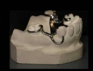
Identify the image?
Correct
ANSWER
I Bar claspOTHER OPTIONS
• NILSYNOPSIS
• It is a retentive clasp used in removable partial dentures.
• The I-bar approaches the undercut from a gingival direction.
• It looks like a straight or slightly curved bar that extends from the base or meshwork and contacts the tooth surface at a point near the gingival third of the crown.
• It resembles the capital letter ‘I’ when viewed from the side.REFERENCE
Stewart’s Clinical Removable Partial Denture ,4th editionIncorrect
ANSWER
I Bar claspOTHER OPTIONS
• NILSYNOPSIS
• It is a retentive clasp used in removable partial dentures.
• The I-bar approaches the undercut from a gingival direction.
• It looks like a straight or slightly curved bar that extends from the base or meshwork and contacts the tooth surface at a point near the gingival third of the crown.
• It resembles the capital letter ‘I’ when viewed from the side.REFERENCE
Stewart’s Clinical Removable Partial Denture ,4th edition -
Question 101 of 150
101. Question
A patient came to your clinic with pain in TMJ and tenderness of muscle with difficulty in swallowing after insertion of complete denture. What could be the possible reason?
Correct
ANSWER
Increased vertical dimension causes clicking, pain, and TMJ problemsOTHER OPTIONS
Refer SynopsisSYNOPSIS
Effects of increased vertical dimension
• Increased trauma – discomfort
• Increased lower- facial height – loss of free-way space
• Cheek biting
• Difficulty in swallowing and speech
• Pain and clicking in the temporomandibular joint
• Stretching of facial muscles – appearance
• Residual ridge resorption
• Drooling of saliva
• Long-term use of complete dentures with increased vertical dimension can cause TMJ disorders.REFERENCE
Post Insertion Problems And Their Management In Complete Denture Journal of Evolution of Medical and Dental Sciences.Vol 2(3) Jan 2013 pp 194-99Incorrect
ANSWER
Increased vertical dimension causes clicking, pain, and TMJ problemsOTHER OPTIONS
Refer SynopsisSYNOPSIS
Effects of increased vertical dimension
• Increased trauma – discomfort
• Increased lower- facial height – loss of free-way space
• Cheek biting
• Difficulty in swallowing and speech
• Pain and clicking in the temporomandibular joint
• Stretching of facial muscles – appearance
• Residual ridge resorption
• Drooling of saliva
• Long-term use of complete dentures with increased vertical dimension can cause TMJ disorders.REFERENCE
Post Insertion Problems And Their Management In Complete Denture Journal of Evolution of Medical and Dental Sciences.Vol 2(3) Jan 2013 pp 194-99 -
Question 102 of 150
102. Question
If relining and rebasing are done on the denture, what will happen?
Correct
ANSWER
Makes the denture more stable without changing the denture teethOTHER OPTION
• Not applicableSYNOPSIS
• Relining is done simply to improve the denture’s fitting.
• Rebasing is a more dramatic adjustment that entails retrofitting the dentures by replacing the entire acrylic base with new acrylic. The procedure makes the denture more stable without changing the denture teeth.REFERENCE
Denture Repair How a Dentist Can Rebase Your Dentures. Randal SIncorrect
ANSWER
Makes the denture more stable without changing the denture teethOTHER OPTION
• Not applicableSYNOPSIS
• Relining is done simply to improve the denture’s fitting.
• Rebasing is a more dramatic adjustment that entails retrofitting the dentures by replacing the entire acrylic base with new acrylic. The procedure makes the denture more stable without changing the denture teeth.REFERENCE
Denture Repair How a Dentist Can Rebase Your Dentures. Randal S -
Question 103 of 150
103. Question
A 40-year-old patient presents with a purplish-red, sessile growth on the gingiva near the mandibular anterior teeth. Histopathological analysis reveals multinucleated giant cells within a fibrovascular stroma. What is the most appropriate treatment for the condition?
Correct
ANSWER
Complete surgical excision with curettage of underlying boneOTHER OPTIONS
• Not applicableSYNOPSIS
• The histology confirms multinucleated giant cells in a vascular stroma indicative of peripheral giant cell granuloma.
• PGCG requires complete surgical excision, including curettage of the underlying bone, to reduce recurrence risk.REFERENCE
Neville, B. W., Damm, D. D., Allen, C. M., & Chi, A. C. (2015). Oral and Maxillofacial Pathology.Incorrect
ANSWER
Complete surgical excision with curettage of underlying boneOTHER OPTIONS
• Not applicableSYNOPSIS
• The histology confirms multinucleated giant cells in a vascular stroma indicative of peripheral giant cell granuloma.
• PGCG requires complete surgical excision, including curettage of the underlying bone, to reduce recurrence risk.REFERENCE
Neville, B. W., Damm, D. D., Allen, C. M., & Chi, A. C. (2015). Oral and Maxillofacial Pathology. -
Question 104 of 150
104. Question
In pregnant patients, what changes occur with her gingiva?
Correct
ANSWER
Increase in gingival fluid and bacteriaOTHER OPTIONS
• Not applicableSYNOPSIS
• Pregnant women have increased gingival fluid and increased growth of bacteria.
• A rise in the ovarian hormones, estrogen and progesterone, increases the risk of inflammation of the gingiva leading to rise in GCF.
• Both pregnancy and use of oral contraceptives (OCs) increases the levels of these hormones. These levels stimulate bacterial growth in the mouth, resulting in a shift in bacteria flora.REFERENCE
Carranza 11th edition chapter 27Incorrect
ANSWER
Increase in gingival fluid and bacteriaOTHER OPTIONS
• Not applicableSYNOPSIS
• Pregnant women have increased gingival fluid and increased growth of bacteria.
• A rise in the ovarian hormones, estrogen and progesterone, increases the risk of inflammation of the gingiva leading to rise in GCF.
• Both pregnancy and use of oral contraceptives (OCs) increases the levels of these hormones. These levels stimulate bacterial growth in the mouth, resulting in a shift in bacteria flora.REFERENCE
Carranza 11th edition chapter 27 -
Question 105 of 150
105. Question
A patient presents with a clean, 1 cm laceration on the forearm sustained during a household activity. Tetanus vaccination is up to date.What is the most appropriate management of this wound?
Correct
ANSWER
Irrigate the wound with saline and close it with suturesOTHER OPTIONS
• Refer SynopsisSYNOPSIS
• Clean wounds can be irrigated with saline and closed primarily if tetanus vaccination is up to date.
• There is no need for immunoglobulin or delayed closure.REFERENCE
Fitzpatrick, T. B., et al. (2017). Emergency Medicine – Clinical Essentials.Incorrect
ANSWER
Irrigate the wound with saline and close it with suturesOTHER OPTIONS
• Refer SynopsisSYNOPSIS
• Clean wounds can be irrigated with saline and closed primarily if tetanus vaccination is up to date.
• There is no need for immunoglobulin or delayed closure.REFERENCE
Fitzpatrick, T. B., et al. (2017). Emergency Medicine – Clinical Essentials. -
Question 106 of 150
106. Question
A 48-year-old renal transplant patient presents with a white, non-wipeable patch on the lateral border of the tongue. The patient is on long-term immunosuppressants. Biopsy confirms oral hairy leukoplakia.What is the most appropriate management for this lesion?
Correct
ANSWER
Monitor the lesion and manage underlying immunosuppressionOTHER OPTIONS
• Not applicableSYNOPSIS
• Oral hairy leukoplakia is caused by Epstein-Barr virus and is associated with immunosuppression.
• Management involves addressing the underlying immunosuppressive state rather than direct treatment of the lesion, as it is benign.REFERENCE
Neville, B. W., Damm, D. D., Allen, C. M., & Chi, A. C. (2015). Oral and Maxillofacial Pathology.Incorrect
ANSWER
Monitor the lesion and manage underlying immunosuppressionOTHER OPTIONS
• Not applicableSYNOPSIS
• Oral hairy leukoplakia is caused by Epstein-Barr virus and is associated with immunosuppression.
• Management involves addressing the underlying immunosuppressive state rather than direct treatment of the lesion, as it is benign.REFERENCE
Neville, B. W., Damm, D. D., Allen, C. M., & Chi, A. C. (2015). Oral and Maxillofacial Pathology. -
Question 107 of 150
107. Question
A 30-year-old patient with a history of Crohn’s disease presents with painful ulcers on the buccal mucosa. Clinical examination shows irregular, erythematous borders with surrounding inflammation.What is the most appropriate management of these oral lesions?
Correct
ANSWER
Administer topical corticosteroids and ensure IBD controlOTHER OPTIONS
• Not applicableSYNOPSIS
Crohn’s disease can present oral lesions, such as aphthous ulcers, cobblestone mucosa, or diffuse swelling. These lesions are inflammatory, and topical corticosteroids (e.g., triamcinolone acetonide) are the first line of treatment
• Oral ulcers in inflammatory bowel disease (IBD) are treated with topical corticosteroids to alleviate symptoms.
• Optimal management of Crohn’s disease is crucial to reduce recurrence.REFERENCE
Chapple, I. L., et al. (2019). Oral manifestations of systemic diseases. Periodontology 2000.Incorrect
ANSWER
Administer topical corticosteroids and ensure IBD controlOTHER OPTIONS
• Not applicableSYNOPSIS
Crohn’s disease can present oral lesions, such as aphthous ulcers, cobblestone mucosa, or diffuse swelling. These lesions are inflammatory, and topical corticosteroids (e.g., triamcinolone acetonide) are the first line of treatment
• Oral ulcers in inflammatory bowel disease (IBD) are treated with topical corticosteroids to alleviate symptoms.
• Optimal management of Crohn’s disease is crucial to reduce recurrence.REFERENCE
Chapple, I. L., et al. (2019). Oral manifestations of systemic diseases. Periodontology 2000. -
Question 108 of 150
108. Question
A 12-year-old child presents with a hyperdivergent skeletal pattern, increased lower anterior facial height, and a Class II malocclusion.Which headgear is most suitable for managing this condition?
Correct
ANSWER
High pull headgearOTHER OPTIONS
• Cervical pull headgear – Cervical pull headgear encourages vertical growth, making it unsuitable for hyperdivergent cases.
• Reverse pull headgear – Reverse pull headgear is used for Class III malocclusion.
• Straight pull headgear – Straight pull headgear applies force parallel to the occlusal plane, which is less effective for hyperdivergent cases.SYNOPSIS
• High-pull headgear applies forces to control vertical growth and reduce lower anterior facial height, making it suitable for hyper divergent cases.REFERENCE
Graber, T. M., Vanarsdall, R. L., & Vig, K. W. (2016). Orthodontics – Current Principles and Techniques.Incorrect
ANSWER
High pull headgearOTHER OPTIONS
• Cervical pull headgear – Cervical pull headgear encourages vertical growth, making it unsuitable for hyperdivergent cases.
• Reverse pull headgear – Reverse pull headgear is used for Class III malocclusion.
• Straight pull headgear – Straight pull headgear applies force parallel to the occlusal plane, which is less effective for hyperdivergent cases.SYNOPSIS
• High-pull headgear applies forces to control vertical growth and reduce lower anterior facial height, making it suitable for hyper divergent cases.REFERENCE
Graber, T. M., Vanarsdall, R. L., & Vig, K. W. (2016). Orthodontics – Current Principles and Techniques. -
Question 109 of 150
109. Question
A 45 years old female patient presented with sudden, severe facial pain triggered by touching the face, chewing or speaking.What may be the diagnosis?
Correct
ANSWER
Trigeminal NeuralgiaOTHER OPTIONS
• TMJ Disorder – can cause pain in your jaw joint and in the muscles that control jaw movement
• Postherpetic neuralgia – Postherpetic neuralgia is the most common complication of shingles. The condition affects nerve fibers and skin, causing burning pain that lasts long after the rash and blisters of shingles disappear.
• Cluster headache – strikes quickly, usually without warning, although you might first have migraine-like nausea and aura. Common signs and symptoms during a headache include: Excruciating pain that is generally situated in, behind or around one eye, but may radiate to other areas of your face, head, and neck.SYNOPSIS
• Trigeminal neuralgia, also called tic douloureux, is a chronic pain condition that affects the trigeminal or 5th cranial nerve.
• TN is a form of neuropathic pain (pain associated with nerve injury or nerve lesion.)
• The typical or classic form of the disorder (called Type 1 or TN1) causes extreme, sporadic, sudden burning or shock-like facial pain that lasts anywhere from a few seconds to as long as two minutes per episode.
• The atypical form of the disorder (called Type 2 or TN2), is characterized by constant aching, burning, stabbing pain of somewhat lower intensity than Type 1. Both forms of pain may occur in the same person, sometimes simultaneously. The intensity of pain can be physically and mentally incapacitating.
• Pain varies, depending on the type of TN, and may range from sudden, severe, and stabbing to a more constant, aching, burning sensation. The intense flashes of pain can be triggered by vibration or contact with the cheek (such as when shaving, washing the face, or applying makeup), brushing teeth, eating, drinking, talking, or being exposed to the wind. The pain may affect a small area of the face or may spread. Bouts of pain rarely occur at night, when the affected individual is sleeping.
• Treatment includes :
– Anticonvulsant medicines-used to block nerve firing are generally effective in treating TN1 but often less effective in TN2. These drugs include carbamazepine, oxcarbazepine, topiramate, gabapentin, pregabalin, clonazepam, phenytoin, lamotrigine, and valproic acid.
– Tricyclic antidepressants such as amitriptyline or nortriptyline can be used to treat pain. Common analgesics and opioids are not usually helpful in treating the sharp, recurring pain caused by TN1, although some individuals with TN2 do respond to opioids. Eventually, if medication fails to relieve pain or produces intolerable side effects such as cognitive disturbances, memory loss, excess fatigue, bone marrow suppression, or allergy, then surgical treatment may be indicated. Since TN is a progressive disorder that often becomes resistant to medication over time, individuals often seek surgical treatment.REFERENCE
Trigeminal Neuralgia Fact Sheet – National Institute of Neurological Disorders and StrokeIncorrect
ANSWER
Trigeminal NeuralgiaOTHER OPTIONS
• TMJ Disorder – can cause pain in your jaw joint and in the muscles that control jaw movement
• Postherpetic neuralgia – Postherpetic neuralgia is the most common complication of shingles. The condition affects nerve fibers and skin, causing burning pain that lasts long after the rash and blisters of shingles disappear.
• Cluster headache – strikes quickly, usually without warning, although you might first have migraine-like nausea and aura. Common signs and symptoms during a headache include: Excruciating pain that is generally situated in, behind or around one eye, but may radiate to other areas of your face, head, and neck.SYNOPSIS
• Trigeminal neuralgia, also called tic douloureux, is a chronic pain condition that affects the trigeminal or 5th cranial nerve.
• TN is a form of neuropathic pain (pain associated with nerve injury or nerve lesion.)
• The typical or classic form of the disorder (called Type 1 or TN1) causes extreme, sporadic, sudden burning or shock-like facial pain that lasts anywhere from a few seconds to as long as two minutes per episode.
• The atypical form of the disorder (called Type 2 or TN2), is characterized by constant aching, burning, stabbing pain of somewhat lower intensity than Type 1. Both forms of pain may occur in the same person, sometimes simultaneously. The intensity of pain can be physically and mentally incapacitating.
• Pain varies, depending on the type of TN, and may range from sudden, severe, and stabbing to a more constant, aching, burning sensation. The intense flashes of pain can be triggered by vibration or contact with the cheek (such as when shaving, washing the face, or applying makeup), brushing teeth, eating, drinking, talking, or being exposed to the wind. The pain may affect a small area of the face or may spread. Bouts of pain rarely occur at night, when the affected individual is sleeping.
• Treatment includes :
– Anticonvulsant medicines-used to block nerve firing are generally effective in treating TN1 but often less effective in TN2. These drugs include carbamazepine, oxcarbazepine, topiramate, gabapentin, pregabalin, clonazepam, phenytoin, lamotrigine, and valproic acid.
– Tricyclic antidepressants such as amitriptyline or nortriptyline can be used to treat pain. Common analgesics and opioids are not usually helpful in treating the sharp, recurring pain caused by TN1, although some individuals with TN2 do respond to opioids. Eventually, if medication fails to relieve pain or produces intolerable side effects such as cognitive disturbances, memory loss, excess fatigue, bone marrow suppression, or allergy, then surgical treatment may be indicated. Since TN is a progressive disorder that often becomes resistant to medication over time, individuals often seek surgical treatment.REFERENCE
Trigeminal Neuralgia Fact Sheet – National Institute of Neurological Disorders and Stroke -
Question 110 of 150
110. Question
What is the use of cleoid discoid instrument in endodontics?
Correct
ANSWER
Both A and BOTHER OPTIONS
• Not applicableSYNOPSIS
• Cleoid Discoid carvers are dental instruments used as part of restorative procedures.
• This instrument is used for trimming excess filling materials.
• The Cleoid Discoid instrument is made of high-grade stainless steel and is used to carve amalgam or wax following natural anatomical form.
• It is an essential carving instrument to develop grooves and triangular ridges.REFERENCE
Basic Guide to Dental Instruments – Carmen Scheller- SheridanIncorrect
ANSWER
Both A and BOTHER OPTIONS
• Not applicableSYNOPSIS
• Cleoid Discoid carvers are dental instruments used as part of restorative procedures.
• This instrument is used for trimming excess filling materials.
• The Cleoid Discoid instrument is made of high-grade stainless steel and is used to carve amalgam or wax following natural anatomical form.
• It is an essential carving instrument to develop grooves and triangular ridges.REFERENCE
Basic Guide to Dental Instruments – Carmen Scheller- Sheridan -
Question 111 of 150
111. Question
Epithelial lining of oral cavity?
Correct
ANSWER
Stratified squamous epitheliumOTHER OPTIONS
• Ciliated columnar epithelium – Ciliated columnar epithelial cells are seen in respiratory epithelial lining.
• Non keratinized squamous epithelium – Junctional epithelium is non keratinised squamous epithelium.
• Stratified ciliated columnar epithelium – Stratified ciliated columnar epithelium lines parts of the respiratory passages, the vas deferens, and the epididymis.SYNOPSIS
• The oral cavity is lined by a mucous membrane (the oral mucosa) consisting of a stratified squamous epithelium, which may or may not be keratinized, and an underlying connective tissue layer, the lamina propria. The surface is kept moist with mucus produced by the major and numerous minor salivary glands.REFERENCE
Tencate oral histologyIncorrect
ANSWER
Stratified squamous epitheliumOTHER OPTIONS
• Ciliated columnar epithelium – Ciliated columnar epithelial cells are seen in respiratory epithelial lining.
• Non keratinized squamous epithelium – Junctional epithelium is non keratinised squamous epithelium.
• Stratified ciliated columnar epithelium – Stratified ciliated columnar epithelium lines parts of the respiratory passages, the vas deferens, and the epididymis.SYNOPSIS
• The oral cavity is lined by a mucous membrane (the oral mucosa) consisting of a stratified squamous epithelium, which may or may not be keratinized, and an underlying connective tissue layer, the lamina propria. The surface is kept moist with mucus produced by the major and numerous minor salivary glands.REFERENCE
Tencate oral histology -
Question 112 of 150
112. Question
A young patient came to you after a bicycle accident with trauma of the central incisor. EPT showed a negative pulp response. Radiographic examination shows the tooth is having an open apex. What is the next step?
Correct
ANSWER
Observe over timeOTHER OPTIONS
• Extraction – Only done if there is no means of saving the tooth.
• Endodontic management – Done in closed apex cases with necrotic pulp changes.
• Pulpotomy – A pulpotomy is a dental procedure in which the pulp in the crown of a tooth is removed in order to save the tooth from infection or decay.SYNOPSIS
• Electric pulp testing is based on stimulation of sensory nerves and requires and relies on subjective assessments and comments from the patient. These can lead to false-positive and false-negative results.
• Younger or anxious patients can have false-positive results due to psychological factors.
• Likewise, false-negative results are possible in teeth with incomplete apical development, trauma, root canal calcification, periodontal disease, or in patients undergoing orthodontic treatment.
• So it’s always better to wait and observe for some time and evaluate the pulp status of the tooth.REFERENCE
Grossman’s endodontic practice pg 378.Incorrect
ANSWER
Observe over timeOTHER OPTIONS
• Extraction – Only done if there is no means of saving the tooth.
• Endodontic management – Done in closed apex cases with necrotic pulp changes.
• Pulpotomy – A pulpotomy is a dental procedure in which the pulp in the crown of a tooth is removed in order to save the tooth from infection or decay.SYNOPSIS
• Electric pulp testing is based on stimulation of sensory nerves and requires and relies on subjective assessments and comments from the patient. These can lead to false-positive and false-negative results.
• Younger or anxious patients can have false-positive results due to psychological factors.
• Likewise, false-negative results are possible in teeth with incomplete apical development, trauma, root canal calcification, periodontal disease, or in patients undergoing orthodontic treatment.
• So it’s always better to wait and observe for some time and evaluate the pulp status of the tooth.REFERENCE
Grossman’s endodontic practice pg 378. -
Question 113 of 150
113. Question
Why should rubber dam application in a patient during endodontic treatment is important?
Correct
ANSWER
Provides a sterile fieldOTHER OPTIONS
• Refer SynopsisSYNOPSIS
• When excavating a deep carious lesion and risking pulpal exposure, use of the rubber dam is strongly recommended to prevent pulpal contamination from oral fluids.i.e it provides a sterile field.
• Other advantages are –
a. Increased access and visibility.
b. Isolates area.
c. Keeps area dry.
d. Protects patient and operator.
e. Retracts soft tissue
f. Preserves and protects materials.REFERENCE
Mosbys review NBDE part 2 pg 55.Incorrect
ANSWER
Provides a sterile fieldOTHER OPTIONS
• Refer SynopsisSYNOPSIS
• When excavating a deep carious lesion and risking pulpal exposure, use of the rubber dam is strongly recommended to prevent pulpal contamination from oral fluids.i.e it provides a sterile field.
• Other advantages are –
a. Increased access and visibility.
b. Isolates area.
c. Keeps area dry.
d. Protects patient and operator.
e. Retracts soft tissue
f. Preserves and protects materials.REFERENCE
Mosbys review NBDE part 2 pg 55. -
Question 114 of 150
114. Question
Color of normal gingiva is interplay between?
Correct
ANSWER
All the aboveOTHER OPTIONS
• NilSYNOPSIS
• The color of the attached and marginal gingiva is generally described as coral pink, it is produced by the vascular supply, the thickness and degree of keratinization of the epithelium, and the presence of pigment containing cells.
• The color also varies among different persons and appears correlated with cutaneous pigmentation. It is lighter in blond individuals with fair complexions than in swarthy, dark-haired individuals.REFERENCE
Carranza 11th edition chapter 2Incorrect
ANSWER
All the aboveOTHER OPTIONS
• NilSYNOPSIS
• The color of the attached and marginal gingiva is generally described as coral pink, it is produced by the vascular supply, the thickness and degree of keratinization of the epithelium, and the presence of pigment containing cells.
• The color also varies among different persons and appears correlated with cutaneous pigmentation. It is lighter in blond individuals with fair complexions than in swarthy, dark-haired individuals.REFERENCE
Carranza 11th edition chapter 2 -
Question 115 of 150
115. Question
The depth of penetration of the probe in the connective tissue apical to the junctional epithelium in the pocket is?
Correct
ANSWER
0.3 mmOTHER OPTIONS
• Anything more than 0.3mm means a gingival or false pocket.SYNOPSIS
• Pocket formation starts as an inflammatory change in the connective tissue wall of the gingival sulcus.
• The cellular and fluid inflammatory exudate causes degeneration of the surrounding connective tissue, including the gingival fibers. Just apical to the junctional epithelium, collagen fibers are destroyed, and the area is occupied by inflammatory cells and edema.
• 0.3mm (the least amount) of connective tissue is left in a periodontal pocket due to connective tissue degeneration.REFERENCE
Carranza 11th edition chapter 13Incorrect
ANSWER
0.3 mmOTHER OPTIONS
• Anything more than 0.3mm means a gingival or false pocket.SYNOPSIS
• Pocket formation starts as an inflammatory change in the connective tissue wall of the gingival sulcus.
• The cellular and fluid inflammatory exudate causes degeneration of the surrounding connective tissue, including the gingival fibers. Just apical to the junctional epithelium, collagen fibers are destroyed, and the area is occupied by inflammatory cells and edema.
• 0.3mm (the least amount) of connective tissue is left in a periodontal pocket due to connective tissue degeneration.REFERENCE
Carranza 11th edition chapter 13 -
Question 116 of 150
116. Question
What is the procedure in which the alveolar cortical plate or the apical foramen is surgically perforated in order to release accumulated tissue exudate?
Correct
ANSWER
TrephinationOTHER OPTIONS
• Root end resection (apical surgery or apicoectomy) – Surgical removal of the apical portion of the root –
• Saucerisation – It is the surgical excavation of tissue – soft tissue, muscle, bone and teeth which include the wound and its surrounding tissue resulting in a shallow depression to help in the drainage of the Infected wound. Most commonly done in Mandible and less common in Maxilla.SYNOPSIS
• Cortical trephination is defined as the surgical perforation of the alveolar cortical plate or apical foramen to release accumulated tissue exudates.
• Indications
– For patients with severe pain of endodontic origin without intraoral or extraoral swelling.
– When drainage cannot be accomplished through the root canal (e.g., posts, filling material, ledging).
• Technique – Cortical trephination involves exposing the cortical bone, making an opening in the bone, and making a pathway through the cancellous bone to the root end.REFERENCE
1. INGLES ENDODONTICS, Pg 969
2. Mosbys review 2nd edition pg 14.Incorrect
ANSWER
TrephinationOTHER OPTIONS
• Root end resection (apical surgery or apicoectomy) – Surgical removal of the apical portion of the root –
• Saucerisation – It is the surgical excavation of tissue – soft tissue, muscle, bone and teeth which include the wound and its surrounding tissue resulting in a shallow depression to help in the drainage of the Infected wound. Most commonly done in Mandible and less common in Maxilla.SYNOPSIS
• Cortical trephination is defined as the surgical perforation of the alveolar cortical plate or apical foramen to release accumulated tissue exudates.
• Indications
– For patients with severe pain of endodontic origin without intraoral or extraoral swelling.
– When drainage cannot be accomplished through the root canal (e.g., posts, filling material, ledging).
• Technique – Cortical trephination involves exposing the cortical bone, making an opening in the bone, and making a pathway through the cancellous bone to the root end.REFERENCE
1. INGLES ENDODONTICS, Pg 969
2. Mosbys review 2nd edition pg 14. -
Question 117 of 150
117. Question
When you decide to do post and core preparation for the RCT tooth. The core must be?
a. Extend to contrabevel
b. Extend to full length tooth preparation
c. Take the same shape of natural tooth
d. Take shape of preparation abutmentCorrect
ANSWER
a and dOTHER OPTIONS
• Not applicableSYNOPSIS
• The amount of coronal tooth structure, along with the position of the tooth in the arch, will dictate the type of core indicated, whether a prefabricated post or a cast post and core is indicated, and whether a crown is needed.
• One of the challenges of core buildup is the need to create an accurate core design according to the desired dimensions of the abutment.
• The physical dimension of the core must be estimated based on the surrounding and opposing tooth anatomies, or by the use of a putty matrix.
• Ferrule effect using a contra bevel in preparation of core acts as an anti-rotational device and as a positive occlusal seat for the post system.
• Contrabevel is placed using flame-shaped diamond point. Bevel is placed at the junction of core and tooth structure.REFERENCE
Prosthetically Guided Technique for Cast Post-and-Core Fabrication – The CompendiumIncorrect
ANSWER
a and dOTHER OPTIONS
• Not applicableSYNOPSIS
• The amount of coronal tooth structure, along with the position of the tooth in the arch, will dictate the type of core indicated, whether a prefabricated post or a cast post and core is indicated, and whether a crown is needed.
• One of the challenges of core buildup is the need to create an accurate core design according to the desired dimensions of the abutment.
• The physical dimension of the core must be estimated based on the surrounding and opposing tooth anatomies, or by the use of a putty matrix.
• Ferrule effect using a contra bevel in preparation of core acts as an anti-rotational device and as a positive occlusal seat for the post system.
• Contrabevel is placed using flame-shaped diamond point. Bevel is placed at the junction of core and tooth structure.REFERENCE
Prosthetically Guided Technique for Cast Post-and-Core Fabrication – The Compendium -
Question 118 of 150
118. Question
A Patient came with a broken post and core. Clinically no visible tooth structure left. IOPA shows a small amount of remaining dentin (less than 1mm.) What is your management?
Correct
ANSWER
ImplantOTHER OPTIONS
• Refer SynopsisSYNOPSIS
• The post should be surrounded by a minimum of 1 mm of sound dentin.
• Clinically no tooth structure is left and only a small amount of dentin is seen in the IOPA, suggesting extraction and implant procedure later.REFERENCE
Grossman Endodontic practice Pg 365.Incorrect
ANSWER
ImplantOTHER OPTIONS
• Refer SynopsisSYNOPSIS
• The post should be surrounded by a minimum of 1 mm of sound dentin.
• Clinically no tooth structure is left and only a small amount of dentin is seen in the IOPA, suggesting extraction and implant procedure later.REFERENCE
Grossman Endodontic practice Pg 365. -
Question 119 of 150
119. Question
Disto lingual extension of mandibular impression will be decided by which muscle?
Correct
ANSWER
Protruding movement of the tongue activates the superior constrictor muscle which molds the disto lingual border of the dentureOTHER OPTIONS
• Refer SynopsisSYNOPSIS
• Border molding in lingual aspect of the lower denture can be divided into three regions
A. Premylohyoid fossa – Influenced by genioglossus muscle.
B. Mylohyoid region – Influenced by mylohyoid muscle.
C. Retromylohyoid curtain – influenced by the superior constrictor the of pharynx and medial pterygoid
• Patient is asked to close the mouth as the dentist applies a downward force on the impression tray.
• When the patient closes his jaw, the medial pterygoid contracts against the superior constrictor of pharynx which is immediately anterior to it.
• Thus the superior constrictor is pushed anteriorly by the contracting medial pterygoid which molds the border of the mandibular denture in the region of the retromylohyoid fossa.
• Protruding the tongue followed by swallowing activates the mylohyoid muscle which raises the floor of the mouth. This helps us determine the length and slope of the lingual flange in the molar region.
• Lingual flange must slope towards the tongue, more or less parallel to the direction of the fibers of the mylohyoid muscle.REFERENCE
Winkler’s Essentials of Complete Denture ProsthodonticsIncorrect
ANSWER
Protruding movement of the tongue activates the superior constrictor muscle which molds the disto lingual border of the dentureOTHER OPTIONS
• Refer SynopsisSYNOPSIS
• Border molding in lingual aspect of the lower denture can be divided into three regions
A. Premylohyoid fossa – Influenced by genioglossus muscle.
B. Mylohyoid region – Influenced by mylohyoid muscle.
C. Retromylohyoid curtain – influenced by the superior constrictor the of pharynx and medial pterygoid
• Patient is asked to close the mouth as the dentist applies a downward force on the impression tray.
• When the patient closes his jaw, the medial pterygoid contracts against the superior constrictor of pharynx which is immediately anterior to it.
• Thus the superior constrictor is pushed anteriorly by the contracting medial pterygoid which molds the border of the mandibular denture in the region of the retromylohyoid fossa.
• Protruding the tongue followed by swallowing activates the mylohyoid muscle which raises the floor of the mouth. This helps us determine the length and slope of the lingual flange in the molar region.
• Lingual flange must slope towards the tongue, more or less parallel to the direction of the fibers of the mylohyoid muscle.REFERENCE
Winkler’s Essentials of Complete Denture Prosthodontics -
Question 120 of 150
120. Question
Histological criteria used to distinguish gingivitis from periodontitis is?
Correct
ANSWER
Howship lacunaeOTHER OPTIONS
• Occlusal trauma and Bone resorption – Howship lacunae and endosteal proliferation are the histological components in this question, whereas trauma from occlusion and bone resorption are clinical components, hence they both are ruled out.
• Endosteal proliferation – Endosteum is a thin vascular membrane of connective tissue that lines the inner surface of the bony tissue that forms the medullary cavity and hence we can safely say it’s not the answer, as gingiva is a soft tissue.SYNOPSIS
• Once inflammation extends from the gingiva to the bone, it spreads to marrow spaces with leukocytic and exudate, new blood vessels, and proliferating fibroblasts, multinuclear osteoclasts and mononuclear phagocytes increase in number and the bone surfaces line up with howships lacunae.
• Howship lacunae is a groove or cavity usually containing osteoclasts that occur in bone that is undergoing resorption.REFERENCE
Carranza 11th edition chapter 14.Incorrect
ANSWER
Howship lacunaeOTHER OPTIONS
• Occlusal trauma and Bone resorption – Howship lacunae and endosteal proliferation are the histological components in this question, whereas trauma from occlusion and bone resorption are clinical components, hence they both are ruled out.
• Endosteal proliferation – Endosteum is a thin vascular membrane of connective tissue that lines the inner surface of the bony tissue that forms the medullary cavity and hence we can safely say it’s not the answer, as gingiva is a soft tissue.SYNOPSIS
• Once inflammation extends from the gingiva to the bone, it spreads to marrow spaces with leukocytic and exudate, new blood vessels, and proliferating fibroblasts, multinuclear osteoclasts and mononuclear phagocytes increase in number and the bone surfaces line up with howships lacunae.
• Howship lacunae is a groove or cavity usually containing osteoclasts that occur in bone that is undergoing resorption.REFERENCE
Carranza 11th edition chapter 14. -
Question 121 of 150
121. Question
Where is the anterior vibrating line located?
Correct
ANSWER
In between the attached tissue of the hard palate and the movable tissue of the soft palateOTHER OPTIONS
• Refer SynopsisSYNOPSIS
• According to Winkler, the Anterior vibration line is an imaginary line at the junction of the attached tissue on the hard palate and the movable tissue next to the soft palate.
• The anterior vibrating line is always on the soft palatal tissues. This should not be confused with the anatomic junction of the hard and soft palate.
• The correctly placed posterior palatal seal will not impinge upon the nondisplaceable tissues of the hard palate.REFERENCE
Winkler S, Essentials of Complete Denture Prosthodontics,pg-107-109Incorrect
ANSWER
In between the attached tissue of the hard palate and the movable tissue of the soft palateOTHER OPTIONS
• Refer SynopsisSYNOPSIS
• According to Winkler, the Anterior vibration line is an imaginary line at the junction of the attached tissue on the hard palate and the movable tissue next to the soft palate.
• The anterior vibrating line is always on the soft palatal tissues. This should not be confused with the anatomic junction of the hard and soft palate.
• The correctly placed posterior palatal seal will not impinge upon the nondisplaceable tissues of the hard palate.REFERENCE
Winkler S, Essentials of Complete Denture Prosthodontics,pg-107-109 -
Question 122 of 150
122. Question
What is an indication of alveoloplasty?
Correct
ANSWER
Eliminates sharp residual ridges and removes bony undercutsOTHER OPTIONS
● Increase the depth of the vestibule – this is achieved by vestibuloplastySYNOPSIS
● Alveoloplasty is a preprosthetic surgical procedure performed to reshape and smoothen the alveolar ridge
● It is indicated for removal of sharp bony projections, irregular ridges, and bony undercuts that interfere with denture fabrication
● The procedure improves denture adaptation, comfort, and stabilityREFERENCE
Peterson’s Principles of Oral and Maxillofacial Surgery – 7th EditionIncorrect
ANSWER
Eliminates sharp residual ridges and removes bony undercutsOTHER OPTIONS
● Increase the depth of the vestibule – this is achieved by vestibuloplastySYNOPSIS
● Alveoloplasty is a preprosthetic surgical procedure performed to reshape and smoothen the alveolar ridge
● It is indicated for removal of sharp bony projections, irregular ridges, and bony undercuts that interfere with denture fabrication
● The procedure improves denture adaptation, comfort, and stabilityREFERENCE
Peterson’s Principles of Oral and Maxillofacial Surgery – 7th Edition -
Question 123 of 150
123. Question
What will be the management of skeletal class III malocclusion due to retrognathic maxilla?
Correct
ANSWER
Lefort advancementOTHER OPTIONS
● Genioplasty – corrects chin deformity, not maxillary deficiencySYNOPSIS
● Skeletal Class III malocclusion due to retrognathic maxilla results from maxillary deficiency rather than mandibular excess
● The treatment of choice is maxillary advancement, commonly performed using Le Fort I osteotomy
● Advancing the maxilla improves facial profile, occlusion, and functional relationship between the jawsREFERENCE
Peterson’s Principles of Oral and Maxillofacial Surgery – 7th EditionIncorrect
ANSWER
Lefort advancementOTHER OPTIONS
● Genioplasty – corrects chin deformity, not maxillary deficiencySYNOPSIS
● Skeletal Class III malocclusion due to retrognathic maxilla results from maxillary deficiency rather than mandibular excess
● The treatment of choice is maxillary advancement, commonly performed using Le Fort I osteotomy
● Advancing the maxilla improves facial profile, occlusion, and functional relationship between the jawsREFERENCE
Peterson’s Principles of Oral and Maxillofacial Surgery – 7th Edition -
Question 124 of 150
124. Question
Patient has been smoking 15 -20 cigarettes a day for 8 years and he comes with a complaint of pain in gingiva, final diagnosis was chronic gingivitis. What is the intensity of inflammation pn this case?
Correct
ANSWER
Less intensity than a non-smokerOTHER OPTIONS
• Not applicableSYNOPSIS
• Clinical signs of inflammation are less pronounced in smokers than in nonsmokers. This may result from alterations in the inflammatory response and microcirculation in smokers.
• Smoking causes alterations in the vascular response of the gingival tissues.
• In addition, the oxygen concentration in healthy gingival tissues appears to be lower in smokers than in nonsmokers.
• Subgingival temperatures are lower in smokers than nonsmokers, and recovery from the vasoconstriction caused by local anesthetic administration takes longer in smokers.REFERENCE
Carranza 11th edition chapter 26Incorrect
ANSWER
Less intensity than a non-smokerOTHER OPTIONS
• Not applicableSYNOPSIS
• Clinical signs of inflammation are less pronounced in smokers than in nonsmokers. This may result from alterations in the inflammatory response and microcirculation in smokers.
• Smoking causes alterations in the vascular response of the gingival tissues.
• In addition, the oxygen concentration in healthy gingival tissues appears to be lower in smokers than in nonsmokers.
• Subgingival temperatures are lower in smokers than nonsmokers, and recovery from the vasoconstriction caused by local anesthetic administration takes longer in smokers.REFERENCE
Carranza 11th edition chapter 26 -
Question 125 of 150
125. Question
Which nerve can be affected during mentoplasty or genioplasty?
Correct
ANSWER
Lower branch of the facial nerveOTHER OPTIONS
● Not applicableSYNOPSIS
● Mentoplasty or genioplasty involves surgical procedures in the chin region for correction of chin deformities
● The marginal mandibular branch (lower branch) of the facial nerve is at risk during surgical dissection in this area
● Injury to this nerve may lead to weakness or asymmetry of the lower lipREFERENCE
Peterson’s Principles of Oral and Maxillofacial Surgery – 7th EditionIncorrect
ANSWER
Lower branch of the facial nerveOTHER OPTIONS
● Not applicableSYNOPSIS
● Mentoplasty or genioplasty involves surgical procedures in the chin region for correction of chin deformities
● The marginal mandibular branch (lower branch) of the facial nerve is at risk during surgical dissection in this area
● Injury to this nerve may lead to weakness or asymmetry of the lower lipREFERENCE
Peterson’s Principles of Oral and Maxillofacial Surgery – 7th Edition -
Question 126 of 150
126. Question
Which instrument is used for disimpacting the maxilla in Le Fort fractures?
Correct
ANSWER
Rowes’ forcepsOTHER OPTIONS
● Refer SynopsisSYNOPSIS
● Rowe’s forceps (or Rowe’s Maxillary Disimpaction Forceps) are the specialized instruments used for disimpacting the maxilla in Le Fort fractures
● These forceps are designed with one straight blade (for the hard palate) and one curved blade (for the nasal floor) to provide a stable, firm grip for moving the maxilla, allowing for strong, controlled, and synchronized downward and forward traction to achieve mobility and proper occlusion.
● Walsham’s forceps and Asche’s forceps are primarily used for the reduction of nasal bone fractures, not for the disimpaction of the entire maxillary complex.REFERENCE
Peterson’s Principles of Oral and Maxillofacial Surgery – 7th EditionIncorrect
ANSWER
Rowes’ forcepsOTHER OPTIONS
● Refer SynopsisSYNOPSIS
● Rowe’s forceps (or Rowe’s Maxillary Disimpaction Forceps) are the specialized instruments used for disimpacting the maxilla in Le Fort fractures
● These forceps are designed with one straight blade (for the hard palate) and one curved blade (for the nasal floor) to provide a stable, firm grip for moving the maxilla, allowing for strong, controlled, and synchronized downward and forward traction to achieve mobility and proper occlusion.
● Walsham’s forceps and Asche’s forceps are primarily used for the reduction of nasal bone fractures, not for the disimpaction of the entire maxillary complex.REFERENCE
Peterson’s Principles of Oral and Maxillofacial Surgery – 7th Edition -
Question 127 of 150
127. Question
A patient reports for follow-up 24 months after replantation of an avulsed tooth. Clinical examination reveals ankylosis without evidence of root resorption. What is the expected future outcome regarding root resorption in such a tooth?
Correct
ANSWER
Root resorption will increaseOTHER OPTIONS
● Root resorption will reduce greatly – Ankylosed replanted teeth generally show progressive replacement resorption over time.SYNOPSIS
● Ankylosis after tooth replantation commonly progresses to replacement root resorption because the root surface becomes fused directly to alveolar bone.
● Over time, normal bone remodeling gradually replaces the root structure with bone, leading to progressive resorption.
● According to the International Association of Dental Traumatology (IADT) guidelines, ankylosed replanted teeth should be monitored long term because replacement resorption is a common sequela, especially after delayed reimplantation.REFERENCE
IADT Guidelines for the Management of Traumatic Dental Injuries – Avulsion of Permanent TeethIncorrect
ANSWER
Root resorption will increaseOTHER OPTIONS
● Root resorption will reduce greatly – Ankylosed replanted teeth generally show progressive replacement resorption over time.SYNOPSIS
● Ankylosis after tooth replantation commonly progresses to replacement root resorption because the root surface becomes fused directly to alveolar bone.
● Over time, normal bone remodeling gradually replaces the root structure with bone, leading to progressive resorption.
● According to the International Association of Dental Traumatology (IADT) guidelines, ankylosed replanted teeth should be monitored long term because replacement resorption is a common sequela, especially after delayed reimplantation.REFERENCE
IADT Guidelines for the Management of Traumatic Dental Injuries – Avulsion of Permanent Teeth -
Question 128 of 150
128. Question
A patient with a replanted avulsed tooth is recalled periodically to assess healing and long-term prognosis. Which of the following is most important for evaluating the prognosis of the replanted tooth?
Correct
ANSWER
Follow-upOTHER OPTIONS
● Flexible wire – Used for splinting the replanted tooth, not for prognosis evaluation.
● Ridge wire – Not a standard method for assessing prognosis of replanted teeth.
● PD wire – Not used for prognostic evaluation in avulsion cases.SYNOPSIS
● Long-term prognosis of a replanted avulsed tooth is determined mainly through periodic clinical and radiographic follow-up.
● Follow-up helps detect complications such as pulpal necrosis, inflammatory root resorption, ankylosis, and replacement resorption.
● According to the International Association of Dental Traumatology (IADT) guidelines, regular follow-up appointments are essential after replantation to evaluate healing and treatment success.REFERENCE
IADT Guidelines for the Management of Traumatic Dental Injuries – Avulsion of Permanent Teeth.Incorrect
ANSWER
Follow-upOTHER OPTIONS
● Flexible wire – Used for splinting the replanted tooth, not for prognosis evaluation.
● Ridge wire – Not a standard method for assessing prognosis of replanted teeth.
● PD wire – Not used for prognostic evaluation in avulsion cases.SYNOPSIS
● Long-term prognosis of a replanted avulsed tooth is determined mainly through periodic clinical and radiographic follow-up.
● Follow-up helps detect complications such as pulpal necrosis, inflammatory root resorption, ankylosis, and replacement resorption.
● According to the International Association of Dental Traumatology (IADT) guidelines, regular follow-up appointments are essential after replantation to evaluate healing and treatment success.REFERENCE
IADT Guidelines for the Management of Traumatic Dental Injuries – Avulsion of Permanent Teeth. -
Question 129 of 150
129. Question
There is mobility in the midface with step deformity in frontozygomatic suture. What is the diagnosis?
Correct
ANSWER
Le Fort III fractureOTHER OPTIONS
● Le Fort I fracture – Produces mobility of the maxillary alveolar segment only without involvement of the frontozygomatic suture.
● Le Fort II fracture – Causes pyramidal midface mobility but usually does not produce step deformity at the frontozygomatic suture.
● Bilateral zygomatic complex fracture – Can cause bilateral frontozygomatic step deformity, but generalized midface mobility is more typical of Le Fort III fracture.SYNOPSIS
● Le Fort III fracture is also called craniofacial dysjunction and involves separation of the entire midface from the cranial base.
● The fracture line traverses the frontozygomatic sutures, leading to palpable step deformity in that region.
● Mobility of the entire midface together with frontozygomatic involvement is therefore a characteristic sign of Le Fort III fracture.REFERENCE
Peterson’s Principles of Oral and Maxillofacial Surgery – 7th EditionIncorrect
ANSWER
Le Fort III fractureOTHER OPTIONS
● Le Fort I fracture – Produces mobility of the maxillary alveolar segment only without involvement of the frontozygomatic suture.
● Le Fort II fracture – Causes pyramidal midface mobility but usually does not produce step deformity at the frontozygomatic suture.
● Bilateral zygomatic complex fracture – Can cause bilateral frontozygomatic step deformity, but generalized midface mobility is more typical of Le Fort III fracture.SYNOPSIS
● Le Fort III fracture is also called craniofacial dysjunction and involves separation of the entire midface from the cranial base.
● The fracture line traverses the frontozygomatic sutures, leading to palpable step deformity in that region.
● Mobility of the entire midface together with frontozygomatic involvement is therefore a characteristic sign of Le Fort III fracture.REFERENCE
Peterson’s Principles of Oral and Maxillofacial Surgery – 7th Edition -
Question 130 of 150
130. Question
In which all conditions open bite is seen?
Correct
ANSWER
All of the aboveOTHER OPTIONS
● Not applicableSYNOPSIS
● An open bite (specifically anterior open bite) is a characteristic finding in all the mentioned conditions due to the loss of posterior vertical height or downward displacement of the maxilla.
● Le Fort II Fractures – These pyramidal fractures result in a floating maxilla, leading to posterior maxillary premature contact and a subsequent anterior open bite.
● Unilateral Condyle Fracture – Displaced subcondylar fractures cause shortening of the ramus height, creating premature contact of the molars on the affected side and a contralateral open bite.
● Bilateral Condyle Fracture: These commonly result in a symmetric anterior open bite due to bilateral shortening of the ramal height.REFERENCE
Peterson’s Principles of Oral and Maxillofacial Surgery – 7th EditionIncorrect
ANSWER
All of the aboveOTHER OPTIONS
● Not applicableSYNOPSIS
● An open bite (specifically anterior open bite) is a characteristic finding in all the mentioned conditions due to the loss of posterior vertical height or downward displacement of the maxilla.
● Le Fort II Fractures – These pyramidal fractures result in a floating maxilla, leading to posterior maxillary premature contact and a subsequent anterior open bite.
● Unilateral Condyle Fracture – Displaced subcondylar fractures cause shortening of the ramus height, creating premature contact of the molars on the affected side and a contralateral open bite.
● Bilateral Condyle Fracture: These commonly result in a symmetric anterior open bite due to bilateral shortening of the ramal height.REFERENCE
Peterson’s Principles of Oral and Maxillofacial Surgery – 7th Edition -
Question 131 of 150
131. Question
What is a sign or symptom of a Concussion luxation injury?
Correct
ANSWER
Tooth with positive percussionOTHER OPTIONS
● Fractured clinical crown – Crown fracture is a hard tissue injury and not a feature of concussion injury.
● Tooth that tests non-vital – Teeth with concussion injuries are usually initially vital, although transient pulpal changes may occur.
● Tooth displaced from the socket – Displacement is seen in luxation injuries such as extrusion or lateral luxation, not concussion.SYNOPSIS
● Concussion is an injury to the tooth-supporting structures without abnormal loosening or displacement of the tooth.
● The characteristic clinical finding is tenderness to percussion, resulting in a positive percussion test.
● Mobility and displacement are absent, and radiographic findings are usually normal.REFERENCE
Peterson’s Principles of Oral and Maxillofacial Surgery – 7th EditionIncorrect
ANSWER
Tooth with positive percussionOTHER OPTIONS
● Fractured clinical crown – Crown fracture is a hard tissue injury and not a feature of concussion injury.
● Tooth that tests non-vital – Teeth with concussion injuries are usually initially vital, although transient pulpal changes may occur.
● Tooth displaced from the socket – Displacement is seen in luxation injuries such as extrusion or lateral luxation, not concussion.SYNOPSIS
● Concussion is an injury to the tooth-supporting structures without abnormal loosening or displacement of the tooth.
● The characteristic clinical finding is tenderness to percussion, resulting in a positive percussion test.
● Mobility and displacement are absent, and radiographic findings are usually normal.REFERENCE
Peterson’s Principles of Oral and Maxillofacial Surgery – 7th Edition -
Question 132 of 150
132. Question
A 50 year old woman, on regular daily doses of steroids for her arthritis has gingivitis and needs superficial scaling. What would be your management?
Correct
ANSWER
No need for supplemental steroidsOTHER OPTIONS
• Not applicableSYNOPSIS
• Evidence indicates that a vast majority of individuals with adrenal insufficiency can receive routine dental treatment without the need for supplemental glucocorticosteroids.
• Patients currently taking corticosteroids generally have enough exogenous and endogenous cortisol to handle routine dental procedures if their usual dose is taken within 2 hours of the planned procedure. Thus, for most patients, supplemental corticosteroid administration is not required when uncomplicated minor surgical procedures, including periodontal surgery, are performed with local anesthesia with or without sedation.
• Individuals who may be at risk for adrenal crisis requiring supplementation include those who are undergoing lengthy, major surgical procedures, those expected to have significant blood loss and those who have extremely low adrenal function.REFERENCE
Carranza 11th edition chapter 37Incorrect
ANSWER
No need for supplemental steroidsOTHER OPTIONS
• Not applicableSYNOPSIS
• Evidence indicates that a vast majority of individuals with adrenal insufficiency can receive routine dental treatment without the need for supplemental glucocorticosteroids.
• Patients currently taking corticosteroids generally have enough exogenous and endogenous cortisol to handle routine dental procedures if their usual dose is taken within 2 hours of the planned procedure. Thus, for most patients, supplemental corticosteroid administration is not required when uncomplicated minor surgical procedures, including periodontal surgery, are performed with local anesthesia with or without sedation.
• Individuals who may be at risk for adrenal crisis requiring supplementation include those who are undergoing lengthy, major surgical procedures, those expected to have significant blood loss and those who have extremely low adrenal function.REFERENCE
Carranza 11th edition chapter 37 -
Question 133 of 150
133. Question
What percentage of bone is composed of inorganic material?
Correct
ANSWER
65%OTHER OPTIONS
● Not applicableSYNOPSIS
● Bone is composed of approximately 65% inorganic material and 35% organic material and water by weight.
● The inorganic component consists mainly of hydroxyapatite crystals containing calcium and phosphate.
● The organic matrix is composed primarily of type I collagen and non-collagenous proteins, which provide tensile strength and flexibility.REFERENCE
Ten Cate’s Oral Histology: Development, Structure, and Function – 10th Edition.Incorrect
ANSWER
65%OTHER OPTIONS
● Not applicableSYNOPSIS
● Bone is composed of approximately 65% inorganic material and 35% organic material and water by weight.
● The inorganic component consists mainly of hydroxyapatite crystals containing calcium and phosphate.
● The organic matrix is composed primarily of type I collagen and non-collagenous proteins, which provide tensile strength and flexibility.REFERENCE
Ten Cate’s Oral Histology: Development, Structure, and Function – 10th Edition. -
Question 134 of 150
134. Question
To minimize the risk of black triangles between teeth, the distance from the contact point to the alveolar bone should be
Correct
ANSWER
Less than 5 mmOTHER OPTIONS
• Refer SynopsisSYNOPSIS
• To minimize the risk of black triangles between teeth, the distance from the contact point to the alveolar bone should be 5 mm or less.
• The iconic study by Tarnow et al. who produced the 5 mm rule, states that when the distance from the contact point to the interproximal osseous crest is 5 mm or less, there is complete fill of the gingival embrasures with an interdental papilla.
• For every 1 mm above 5 mm, the chance of complete fill is progressively reduced by 50 percent.
• For square-shaped teeth with wide contact points, the chances of black triangles is minimal compared with triangular teeth having narrow, more incisally positioned contact points.REFERENCE
Black triangle dilemma and its management in esthetic dentistry – Dental Research Journal 2013 JuneIncorrect
ANSWER
Less than 5 mmOTHER OPTIONS
• Refer SynopsisSYNOPSIS
• To minimize the risk of black triangles between teeth, the distance from the contact point to the alveolar bone should be 5 mm or less.
• The iconic study by Tarnow et al. who produced the 5 mm rule, states that when the distance from the contact point to the interproximal osseous crest is 5 mm or less, there is complete fill of the gingival embrasures with an interdental papilla.
• For every 1 mm above 5 mm, the chance of complete fill is progressively reduced by 50 percent.
• For square-shaped teeth with wide contact points, the chances of black triangles is minimal compared with triangular teeth having narrow, more incisally positioned contact points.REFERENCE
Black triangle dilemma and its management in esthetic dentistry – Dental Research Journal 2013 June -
Question 135 of 150
135. Question
A 21 year old male patient came to your clinic with extrusion of 11 followed by a trauma happened 30 minutes ago. What treatment will you recommended?
Correct
ANSWER
Repositioned into socket with gentle pressure and splintOTHER OPTIONS
Not applicableSYNOPSIS
• Extrusion is partial displacement of tooth from its alveolar socket.
• Extruded teeth should be forced back into socket as soon as possible by means of gentle pressure, after accident and preferably done after anesthetizing the region.
• When extruded teeth are repositioned they are quite likely to be retained for a lifetime.
• Then the tooth should be splinted with a flexible splint for a period of 2 weeks.
• Tooth should be tested for vitality once a month and if non vital, perform RCT.REFERENCE
Grossman’s Endodontic Practise – 13th Edition Page No 434.Incorrect
ANSWER
Repositioned into socket with gentle pressure and splintOTHER OPTIONS
Not applicableSYNOPSIS
• Extrusion is partial displacement of tooth from its alveolar socket.
• Extruded teeth should be forced back into socket as soon as possible by means of gentle pressure, after accident and preferably done after anesthetizing the region.
• When extruded teeth are repositioned they are quite likely to be retained for a lifetime.
• Then the tooth should be splinted with a flexible splint for a period of 2 weeks.
• Tooth should be tested for vitality once a month and if non vital, perform RCT.REFERENCE
Grossman’s Endodontic Practise – 13th Edition Page No 434. -
Question 136 of 150
136. Question
Patient comes to your clinic complaining of gum bleeding. On clinical examination there is loss of gingival stippling, areas of swelling that bleed easily on probing. The pocket depth measured was 4-6mm and no loss of attachment seen. What is the most probable diagnosis?
Correct
ANSWER
Chronic gingivitisOTHER OPTIONS
• Acute gingivitis – Acute gingivitis is typically associated with sudden onset of gum inflammation and bleeding but does not usually show pocket depth of 4-6 mm unless severe inflammation causes pseudo-pocketing.
• Medication-induced gingival enlargement – This condition is characterized by gingival overgrowth (often in response to medications like phenytoin, cyclosporine, or calcium channel blockers) rather than swelling and bleeding due to inflammation.
• ANUG – Characteristic ANUG lesions are punched-out, craterlike depressions at the crest of the interdental papillae, subsequently extending to the marginal gingiva and rarely to the attached gingiva and oral mucosa. The surface of the gingival craters is covered by a gray, pseudomembranous slough, demarcated from the remainder of the gingival mucosa by a pronounced linear erythema.SYNOPSIS
• Chronic gingivitis is slow in onset and of long duration, and is painless, unless complicated by acute or subacute exacerbations.
• Chronic gingivitis is a fluctuating disease in which inflammation persists or resolves and normal areas become inflamed with bleeding on probing, loss of stippling, and swelling.
• Pocket depths of 4-6 mm in this case are likely pseudo-pockets caused by gingival swelling, not actual attachment loss.REFERENCE
Carranza 11th edition chapter 8Incorrect
ANSWER
Chronic gingivitisOTHER OPTIONS
• Acute gingivitis – Acute gingivitis is typically associated with sudden onset of gum inflammation and bleeding but does not usually show pocket depth of 4-6 mm unless severe inflammation causes pseudo-pocketing.
• Medication-induced gingival enlargement – This condition is characterized by gingival overgrowth (often in response to medications like phenytoin, cyclosporine, or calcium channel blockers) rather than swelling and bleeding due to inflammation.
• ANUG – Characteristic ANUG lesions are punched-out, craterlike depressions at the crest of the interdental papillae, subsequently extending to the marginal gingiva and rarely to the attached gingiva and oral mucosa. The surface of the gingival craters is covered by a gray, pseudomembranous slough, demarcated from the remainder of the gingival mucosa by a pronounced linear erythema.SYNOPSIS
• Chronic gingivitis is slow in onset and of long duration, and is painless, unless complicated by acute or subacute exacerbations.
• Chronic gingivitis is a fluctuating disease in which inflammation persists or resolves and normal areas become inflamed with bleeding on probing, loss of stippling, and swelling.
• Pocket depths of 4-6 mm in this case are likely pseudo-pockets caused by gingival swelling, not actual attachment loss.REFERENCE
Carranza 11th edition chapter 8 -
Question 137 of 150
137. Question
A patient with abrasion on the teeth and gingivitis complains of bleeding on brushing. On examination, the patient’s oral hygiene measures show brushing twice and using floss too. What change has to be advised for him for better periodontal health?
Correct
ANSWER
Change the brushing techniqueOTHER OPTIONS
• Not applicableSYNOPSIS
• Changing the brushing technique can improve the condition of this patient
• Bass Method or Sulcular cleaning method is the most accepted and effective method for the removal of dental plaque present adjacent to and underneath the gingival margin
• Indications
– Open interproximal areas
– Cervical areas beneath the height of the contour of enamel
– Exposed root surface
– Recommended for patients with or without periodontal involvement
• Technique
– The bristles are held at a 45-degree angle toward the gum line.
– Very slight pressure and vibratory motions are made so that the bristles go slightly beneath the gum line.
– Only small groups of teeth can be done at a time.
– Once an area is complete, move on to the next set of teeth.REFERENCE
Carranza’s Clinical PeriodontologyIncorrect
ANSWER
Change the brushing techniqueOTHER OPTIONS
• Not applicableSYNOPSIS
• Changing the brushing technique can improve the condition of this patient
• Bass Method or Sulcular cleaning method is the most accepted and effective method for the removal of dental plaque present adjacent to and underneath the gingival margin
• Indications
– Open interproximal areas
– Cervical areas beneath the height of the contour of enamel
– Exposed root surface
– Recommended for patients with or without periodontal involvement
• Technique
– The bristles are held at a 45-degree angle toward the gum line.
– Very slight pressure and vibratory motions are made so that the bristles go slightly beneath the gum line.
– Only small groups of teeth can be done at a time.
– Once an area is complete, move on to the next set of teeth.REFERENCE
Carranza’s Clinical Periodontology -
Question 138 of 150
138. Question
A 25-year-old techie presents with tooth sensitivity and enamel loss on the palatal surfaces of maxillary anterior teeth. The patient admits to frequent consumption of acidic beverages. What type of tooth wear is most likely responsible for these clinical findings?
Correct
ANSWER
ErosionOTHER OPTIONS
• Attrition – Dental attrition is a type of tooth wear caused by tooth-to-tooth contact, resulting in loss of tooth tissue, usually starting at the incisal or occlusal surfaces.
• Abrasion – Abrasion is the wearing away of tooth surface caused by friction or a mechanical process
• Abfraction – Abfraction is the loss of tooth structure from flexural forces.SYNOPSIS
• Erosion is the wearing away of the tooth surface by an acid, which dissolves the enamel and the dentine.
• The causes may include dietary, acid regurgitation, environmental, etc
• The erosion is seen as a light yellow patch on the tooth. This indicates that the enamel has been dissolved and the sensitive dentine under the enamel has been exposed.
• This exposure of the dentine will result in pain from sweet, hot, and cold food and drinks. If the erosion penetrates deeper and reaches the nerves and blood vessels, the pain can become severe.
• Treatment includes
– Treating the underlying medical disorder or disease.
– Changing the patient’s lifestyle to avoid the food or beverage.
– Applying fluoride gels or varnishes to the teeth.
– Drinking milk or using other dairy products.
– Using a neutralizing agent such as antacid tablets.
– Applying dentine bonding agents to areas of exposed dentin.REFERENCE
Shafer’s textbook of Oral PathologyIncorrect
ANSWER
ErosionOTHER OPTIONS
• Attrition – Dental attrition is a type of tooth wear caused by tooth-to-tooth contact, resulting in loss of tooth tissue, usually starting at the incisal or occlusal surfaces.
• Abrasion – Abrasion is the wearing away of tooth surface caused by friction or a mechanical process
• Abfraction – Abfraction is the loss of tooth structure from flexural forces.SYNOPSIS
• Erosion is the wearing away of the tooth surface by an acid, which dissolves the enamel and the dentine.
• The causes may include dietary, acid regurgitation, environmental, etc
• The erosion is seen as a light yellow patch on the tooth. This indicates that the enamel has been dissolved and the sensitive dentine under the enamel has been exposed.
• This exposure of the dentine will result in pain from sweet, hot, and cold food and drinks. If the erosion penetrates deeper and reaches the nerves and blood vessels, the pain can become severe.
• Treatment includes
– Treating the underlying medical disorder or disease.
– Changing the patient’s lifestyle to avoid the food or beverage.
– Applying fluoride gels or varnishes to the teeth.
– Drinking milk or using other dairy products.
– Using a neutralizing agent such as antacid tablets.
– Applying dentine bonding agents to areas of exposed dentin.REFERENCE
Shafer’s textbook of Oral Pathology -
Question 139 of 150
139. Question
A 12-year-old boy was brought to the clinic by his parents for a routine dental check up. On examination, you noticed a deep pit and fissure of newly erupted molars. What will you do to protect the newly erupted molars?
Correct
ANSWER
Apply pit and fissure sealantsOTHER OPTIONS
• NilSYNOPSIS
• The use of pit and fissure sealants provides a physical barrier that inhibits microorganisms and food particles accumulation, preventing caries initiation, and arresting caries progression
• The indications for placing a pit and fissure sealant are as follow
– Pits and fissures of deciduous teeth in children when the tooth, or the patient, is at an increased risk of experiencing caries
– Pits and fissures of permanent teeth in children and adolescents when the tooth, or the patient, is at risk of experiencing caries
– Pits and fissures of permanent teeth in adults when the tooth, or the patient, is at risk of experiencing caries
– Incipient carious lesions (non-cavitated) of pits and fissures in children, adolescents, and adults .
– Pit and fissures of primary and permanent teeth should be considered in children and young people with medical, physical, or intellectual disabilities, mostly when systemic health could be jeopardized by dental disease or the need for dental treatmentREFERENCE
Pit and fissure Sealants – NCBI Book ShelfIncorrect
ANSWER
Apply pit and fissure sealantsOTHER OPTIONS
• NilSYNOPSIS
• The use of pit and fissure sealants provides a physical barrier that inhibits microorganisms and food particles accumulation, preventing caries initiation, and arresting caries progression
• The indications for placing a pit and fissure sealant are as follow
– Pits and fissures of deciduous teeth in children when the tooth, or the patient, is at an increased risk of experiencing caries
– Pits and fissures of permanent teeth in children and adolescents when the tooth, or the patient, is at risk of experiencing caries
– Pits and fissures of permanent teeth in adults when the tooth, or the patient, is at risk of experiencing caries
– Incipient carious lesions (non-cavitated) of pits and fissures in children, adolescents, and adults .
– Pit and fissures of primary and permanent teeth should be considered in children and young people with medical, physical, or intellectual disabilities, mostly when systemic health could be jeopardized by dental disease or the need for dental treatmentREFERENCE
Pit and fissure Sealants – NCBI Book Shelf -
Question 140 of 150
140. Question
Why is a Type B autoclave considered the most suitable choice for sterilizing hollow, porous, and wrapped items in a clinical setting?
Correct
ANSWER
Type B autoclaves use a pre-vacuum phase to remove air, ensuring steam penetration into porous and hollow itemsOTHER OPTIONS
• Not applicableSYNOPSIS
• Type B autoclaves are advanced autoclaves that use a pre-vacuum or post-vacuum phase to effectively remove air from the chamber and items being sterilized.
• This feature ensures complete steam penetration into hollow, porous, and wrapped instruments, which might not be effectively sterilized by other autoclave types like Type N.
• They are particularly suited for medical and dental settings where such instruments are frequently used.REFERENCE
Principles and Methods of Sterilization in Health Sciences (2nd ed.). Springfield: Charles C. Thomas.Incorrect
ANSWER
Type B autoclaves use a pre-vacuum phase to remove air, ensuring steam penetration into porous and hollow itemsOTHER OPTIONS
• Not applicableSYNOPSIS
• Type B autoclaves are advanced autoclaves that use a pre-vacuum or post-vacuum phase to effectively remove air from the chamber and items being sterilized.
• This feature ensures complete steam penetration into hollow, porous, and wrapped instruments, which might not be effectively sterilized by other autoclave types like Type N.
• They are particularly suited for medical and dental settings where such instruments are frequently used.REFERENCE
Principles and Methods of Sterilization in Health Sciences (2nd ed.). Springfield: Charles C. Thomas. -
Question 141 of 150
141. Question
The drug which does not induce gingival enlargement?
Correct
ANSWER
Mefenamic acidOTHER OPTIONS
Esthetically disfiguring overgrowth of gingiva is a significant side effect which may be associated with (Hassell and Hefti 1991, Seymour et al. 1996, Seymour 2006)
• Anticonvulsants (e.g. phenytoin, sodium valproate, etc.)
• Immunosuppressant (e.g. cyclosporine A)
• Calcium channel blocking agents (e.g. nifedipine,verapamil, etc.).SYNOPSIS
• Mefenamic acid is used for the short-term treatment of mild to moderate pain from various conditions.
• It is also used to decrease pain and blood loss from menstrual periods.
• Mefenamic acid is known as a nonsteroidal anti-inflammatory drug (NSAID).REFERENCE
Lindhe 6th edition chapter 19Incorrect
ANSWER
Mefenamic acidOTHER OPTIONS
Esthetically disfiguring overgrowth of gingiva is a significant side effect which may be associated with (Hassell and Hefti 1991, Seymour et al. 1996, Seymour 2006)
• Anticonvulsants (e.g. phenytoin, sodium valproate, etc.)
• Immunosuppressant (e.g. cyclosporine A)
• Calcium channel blocking agents (e.g. nifedipine,verapamil, etc.).SYNOPSIS
• Mefenamic acid is used for the short-term treatment of mild to moderate pain from various conditions.
• It is also used to decrease pain and blood loss from menstrual periods.
• Mefenamic acid is known as a nonsteroidal anti-inflammatory drug (NSAID).REFERENCE
Lindhe 6th edition chapter 19 -
Question 142 of 150
142. Question
What are the types of gingival enlargement?
Correct
ANSWER
All of the aboveOTHER OPTIONS
NilSYNOPSIS
• An increase in size of the gingiva is a common feature of gingival disease. The accepted current terms for this condition are gingival enlargement and gingival overgrowth.
• The many types of gingival enlargement can be classified according to etiologic factors and pathologic changes as follows-
• Inflammatory enlargement
• Drug-induced enlargement
• Enlargements associated with systemic diseases or conditions
• Neoplastic enlargement (gingival tumors)
• False enlargement
• Idiopathic enlargement (hereditary gingival fibromatosis)REFERENCE
Carranza 11th edition chapter 9Incorrect
ANSWER
All of the aboveOTHER OPTIONS
NilSYNOPSIS
• An increase in size of the gingiva is a common feature of gingival disease. The accepted current terms for this condition are gingival enlargement and gingival overgrowth.
• The many types of gingival enlargement can be classified according to etiologic factors and pathologic changes as follows-
• Inflammatory enlargement
• Drug-induced enlargement
• Enlargements associated with systemic diseases or conditions
• Neoplastic enlargement (gingival tumors)
• False enlargement
• Idiopathic enlargement (hereditary gingival fibromatosis)REFERENCE
Carranza 11th edition chapter 9 -
Question 143 of 150
143. Question
A 17 year old man is complaining of enlarged gingiva involving most of his teeth. The medical history revealed that he is epileptic and is being treated with phenytoin.What is the management of drug induced gingival enlargement and inflammation?
Correct
ANSWER
Consult physician and non surgical treatmentOTHER OPTIONS
• Not applicableSYNOPSIS
• Patient taking a drug known to cause gingival enlargement and if gingival enlargement is present, the following things must be done –
– Oral hygiene reinforcement
– Chlorhexidine gluconate rinses
– Scaling and root planing
– Possible drug substitution
– Professional recalls
• First, consideration should be given to the possibility of discontinuing the drug or changing the medication. • These possibilities should be examined with the patient’s physician.
• If any drug substitution is attempted, it is important to allow for a 6 to 12-month period to elapse between discontinuation of the offending drug and the possible resolution of gingival enlargement before a decision to implement surgical treatment is made.
• Alternative medications to the anticonvulsant phenytoin include carbamazepine and valproic acid,
to nifedipine – diltiazem or verapamil and for cyclosporine – tacrolimus.REFERENCE
Carranza 12th edition chapter 58Incorrect
ANSWER
Consult physician and non surgical treatmentOTHER OPTIONS
• Not applicableSYNOPSIS
• Patient taking a drug known to cause gingival enlargement and if gingival enlargement is present, the following things must be done –
– Oral hygiene reinforcement
– Chlorhexidine gluconate rinses
– Scaling and root planing
– Possible drug substitution
– Professional recalls
• First, consideration should be given to the possibility of discontinuing the drug or changing the medication. • These possibilities should be examined with the patient’s physician.
• If any drug substitution is attempted, it is important to allow for a 6 to 12-month period to elapse between discontinuation of the offending drug and the possible resolution of gingival enlargement before a decision to implement surgical treatment is made.
• Alternative medications to the anticonvulsant phenytoin include carbamazepine and valproic acid,
to nifedipine – diltiazem or verapamil and for cyclosporine – tacrolimus.REFERENCE
Carranza 12th edition chapter 58 -
Question 144 of 150
144. Question
A dentist while doing RCT caused an instrument to fracture in the canal. In his attempt to bypass the separated instrument, another instrument tip also got fractured in the canal. How will you manage this case?
Correct
ANSWER
Surgical RCTOTHER OPTION
• Not applicableSYNOPSIS
• Fracture of root canal instruments is one of the most troublesome incidents in endodontic therapy.
• Endodontic instruments rarely separate beyond the apical foramen.
• The fractured segment, always accompanied with bacteria and dentine debris, is a foreign object and might cause inflammation.
• Moreover, patients often regard the fractured segment as a broken needle and suffer psychologically.
• Therefore, an attempt to remove the segment from such cases with a surgical approach is often necessary.
• Before surgery, the precise position and size of the fractured instrument should be understood as well as its relation to the root apex and surrounding anatomic structuresREFERENCE
Surgical Removal of Fractured Endodontic Instrument in the Periapex of Mandibular First Molar. Adil GandevivalaIncorrect
ANSWER
Surgical RCTOTHER OPTION
• Not applicableSYNOPSIS
• Fracture of root canal instruments is one of the most troublesome incidents in endodontic therapy.
• Endodontic instruments rarely separate beyond the apical foramen.
• The fractured segment, always accompanied with bacteria and dentine debris, is a foreign object and might cause inflammation.
• Moreover, patients often regard the fractured segment as a broken needle and suffer psychologically.
• Therefore, an attempt to remove the segment from such cases with a surgical approach is often necessary.
• Before surgery, the precise position and size of the fractured instrument should be understood as well as its relation to the root apex and surrounding anatomic structuresREFERENCE
Surgical Removal of Fractured Endodontic Instrument in the Periapex of Mandibular First Molar. Adil Gandevivala -
Question 145 of 150
145. Question
Informed consent requires that the patient be advised of all of the following except one. Which one is the exception?
Correct
ANSWER
Cost of the procedureOTHER OPTIONS
• Not applicableSYNOPSIS
• Informed consent legally and ethically requires discussing
– The nature of the procedure
– Its risks and benefits
– Alternatives, including doing nothing
– Opportunity for the patient to ask questions and voluntarily agree
• While discussing cost is important for transparency and patient satisfaction, it is not a legal requirement of informed consent.REFERENCE
lInformed Consent Form and Important. Richard A WagnerIncorrect
ANSWER
Cost of the procedureOTHER OPTIONS
• Not applicableSYNOPSIS
• Informed consent legally and ethically requires discussing
– The nature of the procedure
– Its risks and benefits
– Alternatives, including doing nothing
– Opportunity for the patient to ask questions and voluntarily agree
• While discussing cost is important for transparency and patient satisfaction, it is not a legal requirement of informed consent.REFERENCE
lInformed Consent Form and Important. Richard A Wagner -
Question 146 of 150
146. Question
Which bur is used to access the cavity through a ceramo metal crown?
Correct
ANSWER
TransmetalOTHER OPTIONS
• Not applicableSYNOPSIS
• Ceramometal crowns consist of ceramic (porcelain) fused to metal.
• A transmetal bur is specifically designed to cut through both porcelain and metal layers efficiently.
• Diamond burs are good for porcelain but not ideal for metal.
• Carbide burs can cut metal but may fracture or skip on ceramic.
• Stainless steel burs are not used for cutting hard restorative materials.REFERENCE
Endodontic treatment of teeth with PFM crowns: considerations and access techniques – Journal of EndodonticsIncorrect
ANSWER
TransmetalOTHER OPTIONS
• Not applicableSYNOPSIS
• Ceramometal crowns consist of ceramic (porcelain) fused to metal.
• A transmetal bur is specifically designed to cut through both porcelain and metal layers efficiently.
• Diamond burs are good for porcelain but not ideal for metal.
• Carbide burs can cut metal but may fracture or skip on ceramic.
• Stainless steel burs are not used for cutting hard restorative materials.REFERENCE
Endodontic treatment of teeth with PFM crowns: considerations and access techniques – Journal of Endodontics -
Question 147 of 150
147. Question
26-year-old man presents to the dental clinic with acute necrotizing ulcerative gingivitis. The patient had poor oral hygiene, multiple caries and partially erupted lower 3rd molars. Which of the following is referred to as the primary incubation zone in this case?
Correct
ANSWER
Pre-existing chronic gingival diseaseOTHER OPTIONS
• Refer SynopsisSYNOPSIS
• The patient having poor oral hygiene indicates the presence of a long-standing gingivitis.
• NUG can occur in otherwise disease-free mouths or can be superimposed on chronic gingivitis or periodontal pockets.
• There is no mention of loss of attachment or any other periodontal destruction and hence periodontal pocket is ruled out.
• NUG does not superimpose on caries or pericoronal flaps, that is, they do not serve as a primary incubation zone.REFERENCE
Carranza FA, Newman MG. Carranza’s Clinical Periodontology. 13th ed. Elsevier, 2018.Incorrect
ANSWER
Pre-existing chronic gingival diseaseOTHER OPTIONS
• Refer SynopsisSYNOPSIS
• The patient having poor oral hygiene indicates the presence of a long-standing gingivitis.
• NUG can occur in otherwise disease-free mouths or can be superimposed on chronic gingivitis or periodontal pockets.
• There is no mention of loss of attachment or any other periodontal destruction and hence periodontal pocket is ruled out.
• NUG does not superimpose on caries or pericoronal flaps, that is, they do not serve as a primary incubation zone.REFERENCE
Carranza FA, Newman MG. Carranza’s Clinical Periodontology. 13th ed. Elsevier, 2018. -
Question 148 of 150
148. Question
What is the distance between two implants?
Correct
ANSWER
3 mmOTHER OPTION
• NILSYNOPSIS
• According to the international literature, the minimum distance between an implant and natural tooth should be at least 1.5-2mm, and between implant and implant should be at least 3mm, in order to obtain an esthetic and functional result lasting in time.REFERENCE
The easy way to implant placement – How to manage distance By CommunityIncorrect
ANSWER
3 mmOTHER OPTION
• NILSYNOPSIS
• According to the international literature, the minimum distance between an implant and natural tooth should be at least 1.5-2mm, and between implant and implant should be at least 3mm, in order to obtain an esthetic and functional result lasting in time.REFERENCE
The easy way to implant placement – How to manage distance By Community -
Question 149 of 150
149. Question
A 58-year-old patient, presents with partially edentulous mandibular arch missing the first molar. The second molar adjacent to the edentulous space is mesially tilted. The dentist plans to design a removable partial denture and needs to select the most appropriate clasp for the tilted second molar, which will serve as an abutment. Which clasp design is most appropriate for a mesially tilted molar in a removable partial denture to achieve retention and minimize stress on the abutment tooth?
Correct
ANSWER
Ring clasp encircling the tooth with a supporting minor connectorOTHER OPTIONS
• The circumferential clasp – It is unsuitable because it cannot effectively engage the undercut on a tilted tooth.
• The reverse action clasp – It is challenging to fabricate, less aesthetic, and creates potential food traps.
• The I-bar clasp – It is generally used for anterior teeth or premolars and is not ideal for tilted molars.SYNOPSIS
• A mesially tilted molar presents a unique challenge for clasp placement due to altered undercut location and path of insertion. .
• A ring clasp is the ideal choice in this scenario because it encircles the tooth, providing retention while compensating for the tilt.
• Additionally, the ring clasp is supported by a minor connector, which enhances stability and minimizes torque on the abutment.REFERENCE
McCracken, W. L., & Carr, A. B. (2016). McCracken’s Removable Partial Prosthodontics (13th ed.). Elsevier.Incorrect
ANSWER
Ring clasp encircling the tooth with a supporting minor connectorOTHER OPTIONS
• The circumferential clasp – It is unsuitable because it cannot effectively engage the undercut on a tilted tooth.
• The reverse action clasp – It is challenging to fabricate, less aesthetic, and creates potential food traps.
• The I-bar clasp – It is generally used for anterior teeth or premolars and is not ideal for tilted molars.SYNOPSIS
• A mesially tilted molar presents a unique challenge for clasp placement due to altered undercut location and path of insertion. .
• A ring clasp is the ideal choice in this scenario because it encircles the tooth, providing retention while compensating for the tilt.
• Additionally, the ring clasp is supported by a minor connector, which enhances stability and minimizes torque on the abutment.REFERENCE
McCracken, W. L., & Carr, A. B. (2016). McCracken’s Removable Partial Prosthodontics (13th ed.). Elsevier. -
Question 150 of 150
150. Question
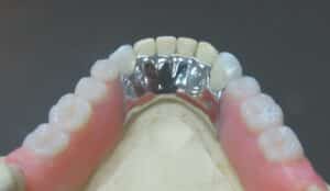
Identify the type of mandibular major connector shown in the image
Correct
ANSWER
Lingual plateOTHER OPTIONS
• Not applicableSYNOPSIS
• Lingual plate
– In cases with anterior tongue thrust or severe bone loss, the lingual plate is used for stabilization.
– Mesial and distal incisal rest seats are prepared on the remaining anterior teeth. These are engaged by metallic extensions (minor connectors) from the lingual plate.
– The lingual plate should fill the interproximal spaces between the weakened teeth to avoid food impaction.
– The plate should have an accurate, thin, knife-edged fit on the lingual aspect of the teeth.
• Functions of lingual plate
– It acts as a major connector for the mandibular prosthesis.
– Provides cross-arch stabilization.
– Provides lingual support for the remaining teeth.REFERENCE
Textbook of Prosthodontics – Deepak NallaswamyIncorrect
ANSWER
Lingual plateOTHER OPTIONS
• Not applicableSYNOPSIS
• Lingual plate
– In cases with anterior tongue thrust or severe bone loss, the lingual plate is used for stabilization.
– Mesial and distal incisal rest seats are prepared on the remaining anterior teeth. These are engaged by metallic extensions (minor connectors) from the lingual plate.
– The lingual plate should fill the interproximal spaces between the weakened teeth to avoid food impaction.
– The plate should have an accurate, thin, knife-edged fit on the lingual aspect of the teeth.
• Functions of lingual plate
– It acts as a major connector for the mandibular prosthesis.
– Provides cross-arch stabilization.
– Provides lingual support for the remaining teeth.REFERENCE
Textbook of Prosthodontics – Deepak Nallaswamy
Last Test Performance Board
Leaderboard: Evergreen Test July - 1
| Pos. | Name | Entered on | Points | Result |
|---|---|---|---|---|
| Table is loading | ||||
| No data available | ||||

Ready to book for the next Evergreen test?
The test will be announced 1 week prior so that you will not miss it.